
Tiroglobulina (Tg)
Liliana M. Bergoglio, Bioquímica Endocrinóloga, Universidad Nacional de Córdoba, Córdoba, Argentina.
E-mail: liberg@uolsinectis.com.ar
Jorge H. Mestman, Médico Endocrinólogo, Universidad del Sur de California, Los Ángeles, CA, Estados Unidos.
NACB: Guía de Consenso para el Diagnóstico y Seguimiento de la Enfermedad Tiroidea
Fuente: Revista Argentina de Endocrinología y Metabilismo, Vol 42, N° 2, Año 2005
Mencionamos con reconocimiento los nombres de los profesionales que participaron en la revisión de la traducción del documento original sobre el cual está basada esta monografía:
Claudio Aranda, Hospital Carlos C. Durand, Buenos Aires, Argentina Aldo H. Coleoni,Universidad Nacional de Córdoba, Córdoba, Argentina
N. Liliana F. de Muñoz, Hospital de Niños de la Santísima Trinidad, Córdoba, Argentina
Silvia Gutiérrez, Hospital Carlos C. Durand, Buenos Aires, Argentina; H. Rubén Harach,Hospital Dr. A. Oñativia, Salta, Argentina
Gustavo C. Maccallini, Hospital Carlos C. Durand, Buenos Aires, Argentina; Mirta B. Miras, Hospital de Niños de la Santísima Trinidad, Córdoba, Argentina
Hugo Niepomniszcze, Universidad Nacional de Buenos Aires, Buenos Aires, Argentina; Adriana Oneto, Hospital Carlos C. Durand, Buenos Aires, Argentina
Eduardo Pusiol, Universidad Nacional de Cuyo, Mendoza, Argentina;Gerardo C. Sartorio, Hospital J. M. Ramos Mejía, Buenos Aires, Argentina
La tiroglobulina (Tg), proteína precursora de la síntesis de las hormonas tiroideas se puede detectar en el suero de la mayoría de los individuos normales si se utiliza un método sensible. El nivel de Tg sérica está influido por tres factores principales: (i) la masa de tejido tiroideo diferenciado presente; (ii) cualquier inflamación o lesión de la glándula tiroides que provoque liberación de Tg; y (iii) el grado de estimulación del receptor de TSH (por TSH, hCG o TRAb). Una concentración elevada de Tg sérica es un indicador no específico de disfunción tiroidea. La mayoría de los pacientes con Tg sérica elevada presentan alteraciones tiroideas benignas. La Tg sérica se utiliza como marcador tumoral en los pacientes con diagnóstico de cáncer diferenciado de tiroides (CDT). Aproximadamente dos tercios de estos presentan un nivel pre-quirúrgico elevado de Tg sérica que confirma la capacidad del tumor de secretar Tg y valida su uso como marcador tumoral post-quirúrgico (307). Por el contrario, cuando la concentración prequirúrgica de Tg sérica no supera los valores normales, no existe evidencia de que el tumor secrete Tg, y un valor post-quirúrgico indetectable es menos tranquilizador. En esos pacientes una concentración post-quirúrgica detectable de Tg sérica podría reflejar la presencia de una importante masa tumoral. De hecho, en general, los cambios post-quirúrgicos representan cambios en la masa tumoral, siempre que se mantenga un nivel constante de TSH mediante terapia con L-T4.
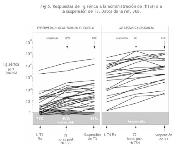
La Tg sérica medida durante el estímulo de TSH (TSH endógena o TSH recombinante humana, rhTSH), es más sensible para la detección del CDT residual o metastásico que una determinación de Tg basal realizada durante el tratamiento con L-T4 (Figura 6) (308). La magnitud del aumento de la Tg sérica en respuesta a la TSH es un indicador de la sensibilidad del tumor a la TSH. Los tumores bien diferenciados típicamente muestran una respuesta estimulada a TSH elevada equivalente a ~10 veces el valor basal de Tg (309). Los tumores escasamente diferenciados que no concentran yoduro pueden presentar una respuesta disminuida al estímulo con TSH (310).
1. Estado Actual de los Métodos de Determinación de Tg
Generalmente, la tiroglobulina se mide en suero, pero también es posible realizar las determinaciones en líquido de quistes tiroideos y/o en material obtenido por punción con aguja fina de masas cervicales no tiroideas (311). La determinación de Tg sérica es técnicamente difícil. En la actualidad, los ensayos inmunométricos (IMA), están superando en popularidad a los métodos por radioinmunoensayo (RIA). La tendencia se debe a que los métodos IMA ofrecen la ventaja práctica de un menor tiempo de incubación, un rango dinámico más amplio y una mayor estabilidad del anticuerpo marcado, y en consecuencia una menor susceptibilidad al daño por marcación que los RIA (312). Hoy los laboratorios pueden escoger entre una serie de métodos IMA tanto isotópicos (inmunoradiométricos, IRMA) como no isotópicos (fundamentalmente quimioluminiscencia, ICMA). No obstante, los métodos IMA suelen ser más susceptibles a interferencias por autoanticuerpos antitiroglobulina (TgAb), que provocan una subestimación de los niveles de Tg sérica. En consecuencia, algunos laboratorios han escogido los métodos RIA para determinarla en pacientes TgAb positivos y restringir el uso de los métodos IMA a los pacientes TgAb negativos. Sin embargo, ningún método puede alegar estar totalmente libre de la interferencia por TgAb, la cual puede provocar resultados inapropiados de Tg.
Además de los problemas con la interferencia de TgAb, los actuales métodos IMA tienen diferencias en la estandarización y en la especificidad, deficiencias en la sensibilidad, precisión interensayo por debajo del nivel óptimo, y potencial predisposición a sufrir efecto “hook” (gancho) a concentraciones elevadas (312).
A. Estandarización
Las concentraciones de Tg sérica determinadas por métodos RIA o IMA presentan marcadas diferencias (312, 313). Un reciente esfuerzo en colaboración patrocinado por la Community Bureau of Reference of the Commission of the European Communities ha desarrollado una nueva Preparación de Referencia Internacional para Tg, CRM-457 (298,314). Este material se puede solicitar al Dr. Christos Profilis, BCR, Rue de la Loi 200, B 1049 Bruselas, Bélgica.
| Recomendación Nº 42. Para los fabricantes que desarrollan métodos para la determinación de Tg |
| *Sería ideal que el diluyente utilizado para los estándares fuese suero humano libre de Tg y TgAb. Se deberían seleccionar matrices no séricas para producir una señal (cuentas radioactivas, unidades relativas de luz, etc.) que sean idénticas al suero humano libre de Tg y TgAb para evitar desvíos (bias) relacionados con las matrices. |
El desvío entre los diferentes métodos de determinación de Tg puede provenir de las diferencias entre las matrices libres de Tg utilizadas para la dilución de los estándares y el suero de pacientes, o diferencias en el reconocimiento de los epitopes por parte de los distintos anticuerpos de Tg usados por los diversos fabricantes. Sería ideal que el diluyente utilizado para los estándares fuese suero humano libre de Tg y TgAb o, como alternativa, una matriz no sérica que hubiera sido seleccionada para producir una señal (cuentas radiactivas, unidades relativas de luz, etc.) que fuese idéntica a la del suero humano libre de Tg y TgAb. Es fundamental que se informe a los médicos antes de que el laboratorio cambie su método para Tg para que puedan realizar una nueva determinación de valores basales en los pacientes con CDT.
La adopción generalizada del estándar CRM-457 se proyectó para reducir, aunque no eliminar la significativa variabilidad inter-método que existe entre los inmunoensayos de Tg de los distintos fabricantes. Se esperaba que la estandarización internacional facilitara un mayor acuerdo entre los diversos estudios publicados y mejorara el uso clínico del seguimiento seriado con Tg en los pacientes con CDT, que a veces tienen determinaciones realizadas en distintos laboratorios. Lamentablemente, el uso del estándar CRM-457 no ha eliminado los problemas de variabilidad entre métodos como se esperaba. En la actualidad, los niveles de Tg sérica determinados por métodos que utilizan la estandarización CRM- 457 pueden presentar diferencias de más de cuatro veces en sus resultados (Figura 7).
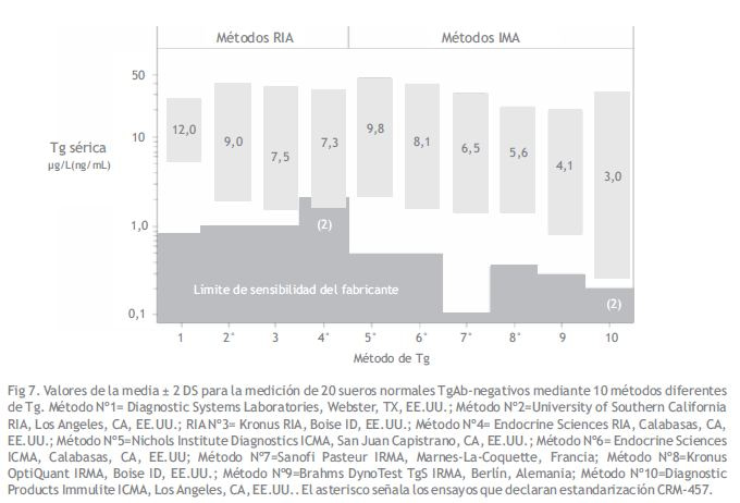
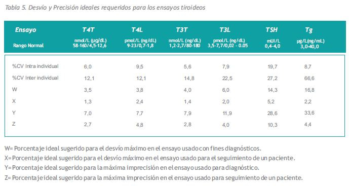
Estas diferencias intermétodos son mayores que la máxima imprecisión aceptada durante el seguimiento de los pacientes individuales (Tabla 5) y excluye el intercambio de diferentes métodos de Tg para el seguimiento a largo plazo de los pacientes con cáncer de tiroides.
| Recomendación Nº 43. Para los laboratorios que consideran cambiar su método de determinación de Tg |
| Seleccionar un método para Tg en función de las características de su comportamiento analítico y no de su costo o conveniencia. Antes de cambiar el método para Tg el laboratorio debería consultar a los médicos que utilizan su método y comparar los resultados entre el método antiguo y el nuevo propuesto utilizando muestras de pacientes TgAb negativos y positivos.*Pacientes TgAb negativos: Si el desvío entre los resultados del método antiguo y el nuevo es > 10%, se debería informar a los médicos y brindarles tiempo suficiente para volver a determinar los valores basales de los pacientes en estado crítico. *Pacientes TgAb positivos: El laboratorio debería advertir a los médicos acerca de la probable dirección de la interferencia en presencia de TgAb. *Si se informan los valores de Tg sérica para las muestras TgAb positivas, se debería incluir una advertencia en cada informe de laboratorio:1.PARA LOS MÉTODOS IMA:Los métodos IMA pueden dar valores de Tg sérica inadecuadamente bajos o indetectables en presencia de TgAb. Los resultados indetectables de Tg sérica no se pueden utilizar como indicadores de ausencia de tumor en un paciente TgAb positivo. Un valor detectable de Tg indica presencia de Tg, pero las concentraciones pueden ser subestimadas.2.PARA LOS MÉTODOS RIA:Los métodos RIA (aunque menos susceptibles a la interferencia) también pueden dar resultados inapropiados de Tg sérica en presencia de TgAb (dependiendo del método). |
B. Sensibilidad
Algunos métodos de Tg carecen de sensibilidad para detectar el límite inferior de referencia eutiroideo que, dependiendo del ensayo, se sitúa aproximadamente en 1-3 ?g/L (ng/mL). Los métodos que no están en condiciones de detectar Tg en todos los sueros normales son insensibles para la búsqueda de recidivas en los pacientes con CDT. Al igual que para TSH, la sensibilidad funcional del ensayo de Tg se determina con el 20% del CV inter-ensayo. El protocolo utilizado para determinar la sensibilidad funcional del ensayo de Tg es el mismo que se describió para TSH (Recomendación Nº 20) con las tres cláusulas descriptas en la Recomendación Nº 44.

| Recomendación Nº 20. Protocolo para obtener la sensibilidad funcional de TSH y el perfil de precisión |
| Medir la TSH en mezclas s de suero humano que cubran el rango del ensayo en por lo menos 10 corridas diferentes. El valor de la mezcla más baja debería estar un 10% por encima del límite de detección y el valor de la mezcla más alta debería estar un 90% por sobre el límite superior del ensayo.*El fenómeno de «arrastre» se debería evaluar analizando primero la mezcla más alta seguida de la más baja. *Utilizar el mismo modo de prueba que para las muestras de pacientes (por ejemplo, simplificado o duplicado). *El operador debería desconocer la presencia de mezclas de sueros de prueba en la corrida. *Las corridas se deberían distribuir en un intervalo clínicamente representativo (por ejemplo 6 a 8 semanas para TSH en pacientes ambulatorios). *Utilizar por lo menos dos lotes diferentes de reactivos y dos calibraciones distintas del instrumento durante el período de prueba. *Cuando se corra el mismo ensayo en dos instrumentos similares, periódicamente se deberían correr duplicados ciegos en cada instrumento para verificar la correlación. |
C. Precisión
La precisión intra- e inter-ensayo, expresada como porcentaje del coeficiente de variación (%CV) es un parámetro importante para la validación del comportamiento analítico de un ensayo de Tg. La precisión se debería determinar utilizando mezclas de sueros TgAb negativos con tres niveles diferentes de Tg (ver Recomendación Nº 44).
La precisión intra-ensayo en los inmunoensayos es mejor que la inter-ensayo, como cabría de esperar. Esto se debe a que las mediciones realizadas dentro de una misma corrida no están sujetas a la variabilidad introducida por el uso de lotes diferentes de reactivos ni por distintas calibraciones de los instrumentos. La precisión intra-ensayo puede ser el parámetro más significativo cuando se evalúa la respuesta de Tg sérica al estímulo con rhTSH (308). En esta prueba, se extrae una muestra basal y una muestra estimulada con rhTSH con 3 a 5 días de intervalo entre sí, y generalmente se determina la Tg de ambas muestras en la misma corrida (Figura 6) (308, 309). En contraste, cuando se utiliza la determinación de Tg para el control seriado, cuanto más prolongado es el intervalo entre corridas mayor la variabilidad y peor la precisión inter-ensayo. Las matrices no humanas utilizadas para determinar la precisión en la zona baja pueden producir una sensibilidad funcional irreal en comparación con las mediciones realizadas con suero humano libre de TgAb como matriz. Es importante que se establezca la sensibilidad funcional y la precisión inter-ensayo con determinaciones distribuidas durante un período entre 6 y 12 meses, ya que éste es el intervalo clínico característico utilizado para el control de los pacientes con CDT.
| Recomendación Nº 44. Sensibilidad funcional y precisión inter-ensayo para los ensayos de Tg |
| La sensibilidad funcional y la precisión interensayo se deberían establecer utilizando el mismo protocolo que para TSH (Recomendación Nº20) con tres consideraciones importantes:*Utilización de mezclas de suero humano que no contengan TgAb, determinados por un inmunoensayo sensible.*Se recomiendan valores óptimos para mezclas de valores bajos, medios y altos: Mezcla de valor bajo (utilizada para determinar la sensibilidad funcional) debería tener un valor de Tg sérica que sea entre 30 y 50 % más elevado que el valor esperado de sensibilidad funcional (SF).[Si SF = 1,0 ug/L (ng/mL) el valor de la mezcla baja debiera ser 1,3 a 1,5 ug/L (ng/mL)] Mezcla de valor medio = ~10 ug/L (ng/mL) es decir, cercano al rango medio normal. Mezcla de valor alto = ~90% del límite superior que informa el fabricante.*El período utilizado para evaluar la precisión inter-ensayo debiera ser por lo menos de 6 meses. Este lapso es más representativo del intervalo clínico utilizado para el control de los pacientes con CDT que el intervalo entre 6 y 8 semanas sugerido para TSH en la Recomendación Nº 20. |
La máxima imprecisión de las determinaciones de Tg sérica sugerida para el seguimiento de pacientes debe ser < 5% (Tabla 5). Es poco probable que los ensayos actuales de Tg puedan mantener una precisión tan estricta a lo largo del intervalo clínicamente relevante entre 6 y 12 meses típicamente utilizados para el control de los pacientes con CDT. Este problema con la precisión se puede superar repitiendo la determinación en muestras previas almacenadas del paciente en la misma corrida que la muestra actual (9).
D. Efecto “hook” a valores elevados
Los métodos IMA se ven afectados por el efecto hook a valores elevados. Los valores falsamente bajos debido a este “efecto gancho” son particularmente problemáticos en los ensayos de marcadores tumorales como la Tg, en donde es frecuente encontrar valores muy elevados cuando los pacientes presentan metástasis avanzada (307, 310, 315). Se produce efecto hook cuando un exceso de antígeno satura la capacidad de unión del anticuerpo de captura. Esto provoca una señal inadecuadamente baja que se traduce en un resultado bajo o paradójicamente normal para un paciente con una concentración excesivamente elevada de Tg sérica (>1000 ?g/L (ng/mL) (312). Los fabricantes de métodos IMA intentan solucionar el problema del efecto hook mediante uno de estos dos procedimientos:
*Diseños de ensayo de dos pasos. Se realiza una primera incubación de la muestra sérica con el anticuerpo de captura antes de que los constituyentes no ligados se eliminen por lavado y se introduzca el anticuerpo marcado, seguido de una segunda incubación.

*Se realizan dos determinaciones (generalmente sin diluir y con dilución 1/10) para cada muestra.
Existe sospecha de efecto “hook” cuando el tubo con la dilución presenta un resultado más alto que la muestra sin diluir. Se realizan más diluciones hasta que el resultado en el tubo con la dilución disminuya y las concentraciones de Tg séricas de dos diluciones consecutivas concuerden.
| Recomendación Nº 45. Detección de efecto «hook» |
| *Se recomienda un diseño de ensayo en dos pasos para minimizar los problemas hook. Los ensayosc»en un paso» que son más propensos al efectoc hook deberían medir cada muestra en dos concentraciones (sin diluir y 1:10) para ver si hay discrepancias entre ambos resultados. *Se debería validar el efecto hook en todos los métodos (de dos pasos o de un paso) antes de su comercialización. *Para verificar el efecto hook, efectuar diluciones 1/10 seriadas de ~ 20 muestras TgAb negativas con concentraciones de Tg sérica superiores a 10.000 ug/L (ng/mL) y ~ 20 muestras TgAb negativas con concentraciones de Tg sérica superiores a 100.000 ug/L (ng/mL) hasta que se demuestre linealidad. |
E. Interferencia por Autoanticuerpos Anti Tiroglobulina (TgAb)
Los autoanticuerpos anti tiroglobulina (TgAb) se detectan con mayor frecuencia en los pacientes con CDT que en la población general (~20 versus ~10%, respectivamente) (276). Las determinaciones seriadas de los TgAb séricos pueden ser indicadores pronósticos independientes de la eficacia del tratamiento o de la recidiva del CDT en los pacientes TgAb positivos (276-278, 316). Cualquier TgAb presente en la muestra tiene el potencial de interferir con un método de Tg (317, 318). Debido a que los TgAb son heterogéneos, ni la medición de la concentración de estos anticuerpos ni un ensayo de recuperación con Tg exógena permiten predecir si los TgAb causarán interferencia (276, 317, 318). Probablemente el signo característico más confiable de la interferencia por TgAb sea la presencia de discordancia entre los RIA y los IMA. La Tg determinada por RIA se caracteriza por valores más elevados que la Tg determinada mediante IMA si la muestra contiene TgAb que provoquen interferencia (276, 309). En la actualidad se ha logrado consenso acerca de que los ensayos de recuperación de Tg no son un método confiable para la detección de TgAb y se los debería eliminar (276, 318).
Los primeros estudios que informaron recuperaciones bajas en ausencia de TgAb en algunos sueros tenían el problema de la insensibilidad de los métodos iniciales para medir TgAb. Cuando se utilizan inmunoensayos cuantitativos con sensibilidad adecuada, los TgAb deberían detectarse siempre cuando la recuperación es baja.
Los métodos inmunométricos no competitivos (IMA) parecen ser más susceptibles a la interferencia producida por TgAb que los RIA, lo que se evidencia por el hallazgo de valores indetectables de Tg en individuos con enfermedad de Graves (319, 318). Aparentemente, en algunos casos los IMA no pueden cuantificar la Tg acomplejada con los TgAb y esta omisión puede provocar subestimación de la concentración de Tg total. Por el contrario los métodos RIA parecen capaces de cuantificar las fracciones de Tg de la muestra tanto libre como ligada a TgAb y característicamente producen valores más altos que los métodos IMA en presencia de TgAb (276, 309). Existe gran variabilidad en la sensibilidad y especificidad de los diferentes ensayos de TgAb. Es esencial que la medición de TgAb sea realizada por el laboratorio que determinará la Tg porque ese laboratorio es responsable de seleccionar el método de TgAb más apropiado para detectar interferencia por TgAb en el método para Tg que utilice.

Cuando se determina Tg en suero que contiene TgAb con métodos RIA e IMA, con frecuencia se observa una discordancia RIA: IMA equivalente a [Tg RIA ? 2 ?g/L (ng/mL): Tg IMA = indetectable]. Esta discordancia parece caracterizar la interferencia por TgAb en una o ambas clases de métodos. Como el umbral actual para una respuesta de Tg positiva estimulada por rhTSH equivale a 2 ?g/L (ng/mL), el grado de discordancia tiene el potencial de influir en la toma de decisiones clínicas (308). Algunos profesionales creen que las mediciones con RIA producen resultados de Tg sérica con mayor validez clínica para los pacientes TgAb positivos que las mediciones con IMA, según se infiere por las correlaciones con el estado clínico y el paralelismo con determinaciones seriadas de TgAb (276, 320). No obstante, cabe destacar que ningún método RIA es inmune a la interferencia por TgAb en todos los sueros TgAb positivos y que la influencia de estos anticuerpos en los diferentes métodos RIA es bastante variable y se relaciona con los componentes del ensayo y las condiciones de incubación. Específicamente, la 125 calidad del trazador I de la Tg, junto con la especificidad del anticuerpo policlonal para Tg determinan la predisposición del método a la interferencia por TgAb (275, 321, 322).
| Recomendación Nº 46. Interferencia por TGAb y ensayos de recuperación |
| *Los ensayos de recuperación no son confiables para la detección de TgAb y se los debería eliminar. Estudios previos que informaron recuperaciones bajas en ausencia de TgAb estaban influenciados por la baja sensibilidad de los métodos iniciales para medir TgAb. Cuando se utilizan inmunoensayos ultrasensibles, siempre es posible detectar TgAb cuando la recuperación es baja. *La discordancia entre las mediciones de Tg realizadas con IMA y RIA en una muestra TgAb positiva sugiere interferencia por TgAb (si los valores habitualmente concuerdan en las muestras TgAb negativas). *Los laboratorios no deberían informar valores indetectables de Tg sérica por método IMA en pacientes TgAb positivos. |
Aunque no existe garantía de que ningún método actual de Tg esté libre de interferencia por TgAb, la subestimación que se produce con la metodología IMA es la dirección de interferencia más grave, ya que este error tiene el potencial de enmascarar la enfermedad metastásica. En consecuencia, los laboratorios no deberían informar valores indetectables de Tg sérica para pacientes TgAb positivos.
2. Determinación de ARN mensajero (ARNm) para Tg
Se ha utilizado la amplificación de ARNm específico de tejido con la reacción en cadena de la polimerasa con transcriptasa reversa (RTPCR) para detectar células cancerígenas circulantes en sangre periférica de los pacientes con melanoma, cáncer de próstata y de mama (326-328). La disponibilidad de cebadores (primers) específicos de Tg permitió la aplicación de esta técnica a la detección de transcriptos de ARNm Tg en sangre. El uso de RT-PCR para detectar recidiva de cáncer tiroideo se informó por primera vez en 1996 (329). A partir de entonces, se ha aplicado la técnica a material de punción de metástasis de ganglios cervicales y se ha visto que es más sensible que la determinación de Tg en el aspirado (330).
Todavía tiene que establecerse el valor clínico de la determinación de ARNm Tg en sangre periférica. Antes de que se pueda utilizar este método para la toma de decisiones terapéuticas en el CDT, es necesario resolver ciertas cuestiones sobre la sensibilidad y especificidad tisular del ARNm Tg en sangre periférica (323- 325).
Varios grupos han desarrollado métodos cuantitativos de RT-PCR para la detección de transcriptos de ARNm Tg en sangre (323-325, 331-333). Estos estudios generalmente encuentran ARNm Tg detectable en todos los individuos normales pero presentan una correlación pobre con la Tg sérica determinada por inmunoensayo (331, 332). También hay diferencias en la correlación entre ARNm Tg y masa tumoral. Algunos estudios han informado que la cantidad de ARNm Tg se correlaciona con la presencia o ausencia de metástasis mientras que otros no informan dicha correlación (324, 331, 333). Es probable que estas discrepancias reflejen diferencias (a) en la sensibilidad y en la especificidad de los primers de Tg y los sistemas RT-PCR, (b) en la sensibilidad de las técnicas de imágenes y de los inmunoensayos de Tg utilizados y (c) en el nivel de TSH del paciente.
Los problemas de especificidad (resultados falsos positivos) constituyen una conocida limitación de la metodología RT-PCR (328, 334).
Se necesitan estudios adicionales para determinar si los niveles detectables de ARNm Tg informados para los pacientes atiróticos sin metástasis conocida reflejan enfermedad clínicamente oculta, artefactos del ensayo o transcripción ilegítima.
Es necesario demostrar la correlación entre los resultados de los ensayos de ARNm Tg y la recidiva clínica, en especial en pacientes ARNm Tg positivos con valores indetectables de Tg sérica antes de que se generalice la adopción del ensayo de ARNm Tg en la práctica clínica. Como este método es más costoso que la determinación de Tg sérica, es probable que si se demuestra que las mediciones de ARNm Tg son útiles para la clínica, se reserven para los pacientes de alto riesgo o TgAb positivos en quienes las determinaciones de Tg sérica no son diagnósticamente confiables.
| Recomendación Nº 47. Para los fabricantes y los laboratorios |
| El folleto con el procedimiento técnico incluido en la caja de reactivos para Tg debería informar sobre las características reales de comportamiento analítico del método (es decir, un comportamiento reproducible en una serie de laboratorios clínicos).*Se deberían estandarizar los ensayos contra la preparación de referencia CRM-457. Los ensayos que no estén estandarizados contra el CRM-457 deberían proveer un factor de corrección.*El valor medio de Tg y los límites de 2 DS del rango de referencia para los individuos normales eutiroideos TgAb negativos (establecidos utilizando la Recomendación Nº48) se deberían citar en todas las publicaciones para permitir la comparación de los valores absolutos. *Los ensayos que no pueden detectar Tg en todos los sueros normales presentan una sensibilidad subóptima para el control de los pacientes con CDT. *Se debería verificar el desvío de la matriz utilizada para la dilución de los estándares (Recomendación Nº 42).*La sensibilidad funcional y la precisión intra- e inter-ensayo se deberían establecer utilizando los protocolos descriptos en la Recomendación N° 44. *La interferencia por TgAb se debería evaluar comprobando las discordancias RIA: IMA en los sueros TgAb positivos [en valores de TgAb de 100 a >1000 kUI/L (UI/mL)]. *Se deberían usar inmunoensayos de sensibilidad adecuada de TgAb, y no ensayos de recuperación con Tg exógena para detectar interferencia por TgAb (ver Recomendación Nº 46). *Los valores de Tg sérica para muestras TgAb positivas no se deberían informar si el método da valores inapropiadamente indetectables en pacientes con CDT con enfermedad documentada. |
3. Valores de Referencia de Tg Sérica
A. Individuos Eutiroideos Normales
Las concentraciones de Tg sérica presentan una distribución normal logarítmica en los individuos eutiroideos. Los valores suelen ser ligeramente más elevados en las mujeres, pero no es necesario establecer rangos de referencia en relación con el género (335). El hábito de fumar es un factor asociado con bocio y valores elevados de Tg sérica (336). Los rangos de referencia de Tg varían según la zona geográfica, ya que reflejan la disponibilidad e ingesta de yoduro (337, 338). La selección de individuos para la cohorte normal para determinar el rango de referencia de Tg debería respetar los siguientes criterios de exclusión:
* Bocio
*Consumo de cigarrillos
*Antecedentes personales o familiares de
enfermedad tiroidea
*Presencia de autoanticuerpos tiroideos (TgAb o
TPOAb)
*TSH sérica 2.0 mUI/L
*Embarazo
B. Valores de Tg sérica después de la cirugía tiroidea
Como se indica en la Recomendación Nº 48, el intervalo de referencia para Tg citado en los informes de laboratorio no corresponde para los pacientes que han sido sometidos a cirugía tiroidea. Durante las primeras semanas después de la cirugía, la Tg sérica estará determinada por la extensión de la intervención, el grado de liberación de Tg debida al daño quirúrgico, y, lo más importante, si el paciente está o no bajo tratamiento con hormona tiroidea. De hecho, la concentración de TSH sérica es un modulador tan potente del nivel de Tg sérica que siempre es necesario conocer el nivel de TSH del paciente antes de establecer el significado de cualquier determinación de Tg sérica.
En las primeras semanas posteriores a la tiroidectomía, se produce una típica disminución de las concentraciones de Tg con una vida media aproximada entre 2 y 4 días, cuando la administración de hormona tiroidea evita el aumento de la TSH (340, 341). En este contexto, la relación entre los valores pre-y post-quirúrgicos (entre 6 y 8 semanas) de Tg puede aportar información que podría influir en el esquema de tratamiento. Durante el seguimiento a largo plazo, las concentraciones de Tg sérica medidas con y sin tratamiento de LT4 (con TSH suprimida o desenfrenada, respectivamente) proporcionan diferente información. La curva de los valores de Tg sérica (con tratamiento con L-T4) es un indicador más específico de un cambio en la masa tumoral que cualquier valor aislado de Tg sérica (122). La concentración de Tg sérica durante el tratamiento con L-T4 es un indicador más estable de masa tumoral que la Tg sérica determinada cuando la TSH está elevada (suspensión de L-T4 o administración de rhTSH) anterior a un rastreo corporal con yodo radioactivo (RAI). Esto se debe a que la magnitud del aumento de Tg sérica estimulada por TSH está influida por el grado y la cronicidad de la elevación de TSH que puede variar de un rastreo a otro. Sin embargo, según lo muestra la Figura 6, como la TSH normalmente estimula más de 10 veces la Tg sérica, las determinaciones de Tg sérica estimuladas por TSH son más sensibles para detectar enfermedad restringida al cuello, que los niveles de Tg sérica determinados durante la supresión de TSH (308, 309). La magnitud de la respuesta de la Tg sérica estimulada por TSH es un indicador de la sensibilidad del tumor a la TSH. Los tumores metastásicos poco diferenciados que son rastreo corporal con yodo radioactivo negativos presentan respuestas disminuidas de Tg estimulada por TSH (310).
| Recomendación Nº 48. Intervalos de referencia para Tg sérica |
| *Los rangos de referencia para Tg se deberían determinar localmente porque las concentraciones de Tg sérica están influenciadas por la ingesta de yoduro: Países con ingesta adecuada de yoduro: El intervalo de referencia para Tg sérica para la población eutiroidea TgAb negativa según los estándares CRM-457 se aproxima los 3 a 40 ?g/L (ng/mL).Países con yododeficiencia: Es posible que se registre un aumento en la media de Tg de la población y del límite superior del rango de referencia relacionado con el grado de carencia de yodo. *Los laboratorios deberían validar su intervalo de referencia para Tg independientemente de los fabricantes. *Se deberían establecer rangos de referencia a partir de los valores logarítmicamente transformados de 120 individuos normales, no fumadores, eutiroideos (TSH 0,5 a 2,0 mUI/L) menores de 40 años sin antecedentes personales ni familiares de enfermedad tiroidea y sin evidencia de TgAb o TPOAb. *Es engañoso citar el rango de referencia normal eutiroideo al informar valores de Tg sérica para los pacientes con CTD tiroidectomizados. Los valores de referencia deberían relacionarse con los límites de referencia eutiroideos para el método, la masa tiroidea y el nivel de TSH.Como ejemplo, los rangos de referencia a continuación serían apropiados para un método de Tg con un rango de referencia eutiroideo de 3-40 ?g/L (ng/mL): Tg ug/L (ng/mL) Condición3 – 40 Glándula tiroides normal (TSH 0,4-4,0 mUI/L)1.5 – 20 Glándula tiroides normal (TSH < 10 Lobectomía tiroidea (TSH < 0,1 mUI/L) < 2 Tiroidectomía casi total (TSH < 0,1 mUI/L) |
4. Usos clínicos de las determinaciones de Tg sérica
La concentración de Tg sérica refleja la masa tiroidea, el daño tiroideo y el estímulo del receptor de TSH (122). En consecuencia, un aumento en la Tg sérica es un hallazgo inespecífico no asociado virtualmente con ninguna patología tiroidea.
A. Patologías no Neoplásicas
Se produce un aumento de la Tg sérica cuando los pacientes tienen bocio y en la mayoría de las patologías hipertiroideas. La concentración baja de Tg sérica puede ser un parámetro útil para la confirmación del diagnóstico de tirotoxicosis facticia o para la investigación de la etiología de hipotiroidismo congénito (342, 343).
La concentración de Tg a veces también es útil,para confirmar la historia pasada de tiroiditis,en la cual la concentración de Tg es habitualmente el último parámetro bioquímico que se normaliza (hasta los 2 años) (344). Estudios recientes sugieren la determinación de Tg sérica como un parámetro para reflejar el estado de yodosuficiencia en una población determinada (337, 338).
| Recomendación Nº 49. Determinación de Tg sérica para patologías no neoplásicas |
| Concentraciones anormalmente altas de Tg resultan de anormalidades en la masa tiroidea, excesiva estimulación tiroidea, daño físico a la tiroides secundario a cirugía, PAAF o tiroiditis. Las determinaciones de Tg sérica son útiles para: *Diagnosticar tirotoxicosis facticia caracterizada por Tg sérica baja. *Investigar la etiología del hipotiroidismo congénito detectado en el screening neonatal. *Evaluar la actividad de la tiroiditis inflamatoria, por ejemplo: tiroiditis subaguda o inducida por amiodarona. |
B. Carcinoma Diferenciado de Tiroides (CDT)
En el contexto del CDT, la concentración de Tg sérica refleja masa tiroidea (tumor o remanente normal), lesión tiroidea (cirugía o PAAF) y estimulación del receptor de TSH (endógena o con rhTSH) (122). Debido a que la TSH es el principal regulador de la concentración de Tg sérica, es difícil interpretar los valores de Tg sin conocer el nivel de TSH del paciente. Aunque no hay un “rango normal de referencia para Tg” para los pacientes con CDT tratados, la relación normal entre la masa tiroidea y la Tg sérica provee un punto de referencia importante. Concretamente, un gramo de tejido tiroideo normal libera ~1 ?g/L (ng/mL) de Tg en la circulación cuando la TSH sérica es normal y ~0,5 ?g/L (ng/mL) cuando se la suprime por debajo de 0,1 mUI/L.
(i) Tg Sérica Pre-quirúrgica
Algunos tumores tiroideos carecen de capacidad para secretar tiroglobulina. En 2/3 de los pacientes con CDT se observa un aumento en el valor pre-quirúrgico de Tg sérica, lo que indica que sus tumores tienen la capacidad de secretar Tg, y por lo tanto el seguimiento postquirúrgico con Tg puede ser de utilidad clínica en ellos. (307). Esta información es fundamental para la interpretación de los resultados post-quirúrgicos de la Tg sérica. Si el nivel pre-quirúrgico está dentro de los límites normales, un valor post-quirúrgico indetectable de Tg sérica es menos tranquilizador porque el tumor pudo ser originariamente no secretor de Tg. La sensibilidad del control postquirúrgico con Tg sérica para la detección de recidiva será mayor cuando el tumor sea relativamente pequeño (
(ii) Determinación de Tg Sérica entre 1 Y 2 meses después de la cirugía Tiroidea
Después de la cirugía tiroidea, las concentraciones de Tg sérica disminuyen rápidamente con una vida media entre ~2 y 4 días (340). La Tg liberada por daño durante la manipulación quirúrgica se debería resolver en gran parte dentro de los primeros dos meses posteriores a la cirugía. Durante este lapso la TSH tendrá una influencia dominante en el nivel de Tg sérica. Si se inicia el tratamiento con hormona tiroidea inmediatamente después de la cirugía para evitar el aumento de TSH, la concentración de Tg sérica declinará a un valor que refleje el tamaño del remanente tiroideo normal más cualquier residuo o metástasis tumoral. Como el remanente tiroideo después de una tiroidectomía casi total habitualmente se aproxima a 2 gramos de tejido, se espera una concentración de Tg sérica equivalente a < 2 ?g/L (ng/mL) cuando la cirugía ha sido exitosa y el nivel de TSH se mantiene por debajo de 0,1 mUI/L.
| Recomendación Nº 50. Determinación de Tg sérica para el carcinoma diferenciado de tiroides (CDT) |
| Pacientes TgAb negativos:*Los valores séricos pre-quirúrgicos (extracción antes o más de 2 semanas después de la PAAF) son útiles para la determinación de la capacidad secretante de Tg del tumor. *La disminución aguda post-quirúrgica de Tg sérica refleja la extensión de la cirugía con una vida media de la Tg entre 3 y 4 días. (Si se administra hormona tiroidea para evitar el aumento de TSH). *No existe «rango normal» para un paciente tiroidectomizado. Los pacientes completamente atireóticos no deben presentar Tg detectable en suero, incluso si la TSH está elevada. *Parámetro útil de referencia: un gramo de tejido tiroideo normal libera ~1 ug/L (ng/mL) de Tg en suero cuando la TSH es normal y ~0,5 ug/L (ng/mL) cuando la TSH está suprimida a < 0,1 mUI/L. *Cuando la Tg sérica es detectable durante el tratamiento con L-T4 (TSH estable) se pueden seguir los cambios en la masa tumoral con determinaciones seriadas de Tg sérica sin interrupción de la hormona tiroidea ni rhTSH. *Cuando la Tg sérica es indetectable bajo tratamiento con L-T4 (y ausencia de TgAb) la Tg sérica estimulada por TSH es más sensible para la detección de enfermedad localizada en el cuello. *Habitualmente se produce un aumento de >5 veces en la Tg sérica con respecto a los valores bajo supresión con LT4 luego del estímulo con TSH (endógena o rhTSH). Estudios comparativos muestran que las respuestas de la Tg estimulada con rhTSH son aproximadamente la mitad que las observadas con TSH endógena siguiendo a la suspensión de la hormona tiroidea.Pacientes TgAb positivos: *Habitualmente presentan respuestas disminuidas o ausentes de Tg sérica estimulada con TSH. *Las determinaciones seriadas de TgAb (por inmunoensayos) son valiosas como marcadores tumorales sustitutos. |
(iii) Determinación de Tg Sérica durante el seguimiento a largo plazo bajo Tratamiento con L-T4.
Cuando el nivel de TSH es estable durante el tratamiento con L-T4, cualquier cambio en el nivel de Tg sérica reflejará un cambio en la masa tumoral. La recidiva clínica en tumores considerados “secretores deficientes de Tg” (valor pre-quirúrgico de Tg en el rango normal) se puede asociar con valores post-quirúrgicos bajos o indetectables de Tg sérica. Por el contrario, la recidiva de tumores considerados “buenos secretores de Tg” (valores prequirúrgicos elevados de Tg) se asocia normalmente con un aumento progresivo en Tg sérica (122).
El perfil de las determinaciones seriadas de Tg sérica, establecido cuando el paciente tiene TSH estable, es más útil clínicamente que un valor aislado de Tg. Sin embargo, es posible interpretar el significado de un valor aislado de Tg conociendo el rango de referencia del ensayo de Tg, la extensión de la cirugía tiroidea y la concentración de TSH (en un estado estable), según lo muestra la Figura 8.
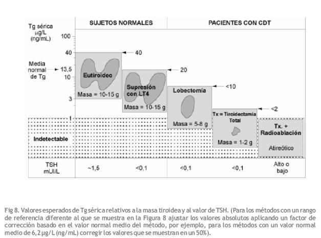
Condiciones Asumidas
*Sin lesión tiroidea reciente (cirugía o PAAF)
*Usando la Recomendación N° 48, la media normal de Tg = 13,5, rango 3-40 (2DS) ?g/L (ng/mL)
*Masa de tejido tiroideo normal = 10-15 gramos
*Un gramo de tejido tiroideo normal produce ~1?g/L (ng/mL) Tg en suero si la TSH es normal
*Un gramo de tejido tiroideo normal produce ~0,5?g/L (ng/mL) Tg si la TSH es < 0.1 mUI/L
(iv) Respuesta de Tg Sérica al estímulo con TSH
La magnitud de la respuesta de la Tg sérica a la TSH endógena (suspensión de la hormona tiroidea) o a la administración de rhTSH es un indicador de la sensibilidad del tumor a la TSH (308, 309). Habitualmente, el estímulo con TSH de los remanentes tiroideos normales o de un tumor bien diferenciado produce un aumento >3 veces de Tg sérica por sobre el nivel basal (con TSH suprimida) en los pacientes TgAb negativos (Figura 6). La respuesta de la Tg sérica a un aumento en la TSH endógena es habitualmente dos veces mayor que con rhTSH (308, 345). Además, los tumores pobremente diferenciados, presentan una respuesta disminuida (< 3 veces) de la Tg sérica al estímulo con TSH (310). Cabe observar que los pacientes TgAb positivos habitualmente presentan una respuesta disminuida o ausente de la Tg al estímulo con rhTSH determinada por la mayoría de los ensayos, incluso cuando la concentración basal de Tg es detectable.
Referencias Bibliográficas
1. Nohr SB, Laurberg P, Borlum KG, Pedersen Km, Johannesen PL, Damm P. Iodine deficiency in pregnancy in Denmark. Regional variations and frequency of individual iodine supplementation. Acta Obstet Gynecol Scand 1993;72:350-3.
2. Glinoer D. Pregnancy and iodine. Thyroid 2001;11:471-81.
3. Hollowell JG, Staehling NW, Hannon WH, Flanders DW, Gunter EW, Maberly GF et al. Iodine nutrition in the Unites States. Trends and public health implications: iodine excretion data from National Health and Nutrition Examination Surveys I and III (1971-1974 and 1988-1994). J Clin Endocrinol Metab 1998;83:3398-400.
4. Wartofsky L, Glinoer D, Solomon d, Nagataki S, Lagasse R, Nagayama Y et al. Differences and similarities in the diagnosis and treatment of Graves disease in Europe, Japan and the United States. Thyroid 1990;1:129-35.
5. Singer PA, Cooper DS, Levy EG, Ladenson PW, Braverman LE, Daniels G et al. Treatment guidelines for patients with hyperthyroidism and hypothyroidism. JAMA 1995;273:808-12.
6. Singer PA, Cooper DS, Daniels GH, Ladenson PW, Greenspan FS, Levy EG et al. Treatment Guidelines for Patients with Thyroid Nodules and Well-differentiated Thyroid Cancer. Arch Intern Med 1996;156:2165-72.
7. Vanderpump MPJ, Ahlquist JAO, Franklyn JA and Clayton RN. Consensus statement for good practice and audit measures in the management of hypothyroidism and hyperthyroidism. Br Med J 1996;313:539-44.
8. Laurberg P, Nygaard B, Glinoer D, Grussendorf M and Orgiazzi J. Guidelines for TSH-receptor antibody measurements in pregnancy: results of an evidence-based symposium organized by the European Thyroid Association. Eur J Endocrinol 1998;139:584-6.
9. Cobin RH, Gharib H, Bergman DA, Clark OH, Cooper DS, Daniels GH et al. AACE/AAES Medical/Surgical Guidelines for Clinical Practice: Management of Thyroid Carcinoma. Endocrine Pract 2001;7:203-20.
10. Ladenson PW, Singer PA, Ain KB, Bagchi N, Bigos ST, Levy EG et al. American Thyroid Association Guidelines for detection of thyroid dysfunction. Arch Intern Med 2000;160:1573-5.
11. Brandi ML, Gagel RJ, Angeli A, Bilezikian JP, Beck-Peccoz P, Bordi C et al. Consensus Guidelines for Diagnosis and Therapy of MEN Type 1 and Type 2. J Clin Endocrinol Metab 2001;86:5658-71.
12. Werner and Ingbar’s “The Thyroid”. A Fundamental and Clinical Text. Lippincott-Raven, Philadelphia 2000. Braverman LE and Utiger RD eds.
13. DeGroot LJ, Larsen PR, Hennemann G, eds. The Thyroid and Its Diseases. (www.thyroidmanager.org) 2000.
14. Piketty ML, D’Herbomez M, Le Guillouzic D, Lebtahi R, Cosson E, Dumont A et al. Clinical comparison of three labeled-antibody immunoassays of free triiodothyronine. Clin Chem 1996;42:933-41.
15. Sapin R, Schlienger JL, Goichot B, Gasser F and Grucker D. Evaluation of the Elecsys free triiodothyronine assay; relevance of age-related reference ranges. Clin Biochem 1998;31:399-404.
16. Robbins J. Thyroid hormone transport proteins and the physiology of hormone binding. In “Hormones in Blood”. Academic Press, London 1996. Gray CH, James VHT, eds. pp 96-110.
17. Demers LM. Thyroid function testing and automation. J Clin Ligand Assay 1999;22:38-41.
18. Hollowell JG, Staehling NW, Hannon WH, Flanders WD, Gunter EW, Spencer CA et al. Serum thyrotropin, thyroxine and thyroid antibodies in the United States population (1988 to 1994): NHANES III. J Clin Endocrinol Metab 2002;87:489-99.
19. Wardle CA, Fraser WD and Squire CR. Pitfalls in the use of thyrotropin concentration as a first-line thyroid-function test. Lancet 2001;357:1013-4.
20. Spencer CA, LoPresti JS, Patel A, Guttler RB, Eigen A, Shen D et al. Applications of a new chemiluminometric thyrotropin assay to subnormal measurement. J Clin Endocrinol Metab 1990;70:453-60.
21. Meikle, A. W., J. D. Stringham, M. G. Woodward and J. C. Nelson. Hereditary and environmental influences on the variation of thyroid hormones in normal male twins. J Clin Endocrinol Metab1 1988;66:588-92.
22. Andersen S, Pedersen KM, Bruun NH and Laurberg P. Narrow individual variations in serum T4 and T3 in normal subjects: a clue to the understanding of subclinical thyroid disease. J Clin Endocrinol Metab 2002;87:1068-72.
23. Cooper, D. S., R. Halpern, L. C. Wood, A. A. Levin and E. V. Ridgway. L-thyroxine therapy in subclinical hypothyroidism. Ann Intern Med 1984;101:18-24.
24. Biondi B, Fazio E, Palmieri EA, Carella C, Panza N, Cittadini A et al. Left ventricular diastolic dysfunction in patients with subclinical hypothyroidism. J Clin Endocrinol Metab 1999;2064-7.
25. Hak AE, Pols HAP, Visser TJ, Drexhage HA, Hofman A and Witteman JCM. Subclinical Hypothyroidism is an independent risk factor for atherosclerosis and myocardial infarction in elderly women: the Rotterdam Study. Ann Intern Med 2000;132:270-8.
26. Michalopoulou G, Alevizaki M, Piperingos G, Mitsibounas D, Mantzos E, Adamopoulos P et al. High serum cholesterol levels in persons with ‘high-normal’ TSH levels: should one extend the definition of subclinical hypothyroidism? Eur J Endocrinol 1998;138:141-5.
27. Beck-Peccoz P, Brucker-Davis F, Persani L, Smallridge RC and Weintraub BD. Thyrotropin-secreting pituitary tumors. Endocrine Rev 1996;17:610-38.
28. Brucker-Davis F, Oldfield EH, Skarulis MC, Doppman JL and Weintraub BD. Thyrotropin-secreting pituitary tumors: diagnostic criteria, thyroid hormone sensitivity and treatment outcome in 25 patients followed at the National Institutes of Health. J Clin Endocrinol Metab 76 1999;:1089-94.
29. Oliveira JH, Persani L, Beck-Peccoz P and Abucham J. Investigating the paradox of hypothyroidism and increased serum thyrotropin (TSH) levels in Sheehan’s syndrome: characterization of TSH carbohydrate content and bioactivity. J Clin Endocrinol Metab 2001;86:1694-9.
30. Uy H, Reasner CA and Samuels MH. Pattern of recovery of the hypothalamic-pituitary thyroid axis following radioactive iodine therapy in patients with Graves’ disease. Amer J Med 1995;99:173-9.
31. Hershman JM, Pekary AE, Berg L, Solomon DH and Sawin CT. Serum thyrotropin and thyroid hormone levels in elderly and middle-aged euthyroid persons. J Am Geriatr Soc 1993;41:823-8.
32. Fraser CG. Age-related changes in laboratory test results. Clinical applications. Drugs Aging 1993;3:246-57.
33. Fraser CG. 2001. Biological Variation: from principles to practice. AACC Press, Washington DC.
34. Drinka PJ, Siebers M and Voeks SK. Poor positive predictive value of low sensitive thyrotropin assay levels for hyperthyroidism in nursing home residents. South Med J 1993;86:1004-7.
35. Vanderpump MPJ, Tunbridge WMG, French JM, Appleton D, Bates D, Rodgers H et al. The incidence of thyroid disorders in the community; a twenty year follow up of the Whickham survey. Clin Endocrinol 1995;43:55-68.
36. Sawin CT, Geller A, Kaplan MM, Bacharach P, Wilson PW, Hershman JM et al. Low serum thyrotropin (thyroid stimulating hormone) in older persons without hyperthyroidism. Arch Intern Med 1991;151:165-8.
37. Parle JV, Maisonneuve P, Sheppard MC, Boyle P and Franklyn JA. Prediction of all-cause and cardiovascular mortality in elderly people from one low serum thyrotropin result: a 10-year study. Lancet 2001;358:861-5.
38. Nelson JC, Clark SJ, Borut DL, Tomei RT and Carlton EI. Age-related changes in serum free thyroxine during childhood and adolescence. J Pediatr 1993;123:899-905.
39. Adams LM, Emery JR, Clark SJ, Carlton EI and Nelson JC. Reference ranges for newer thyroid function tests in premature infants. J Pediatr 1995;126:122-7.
40. Lu FL, Yau KI, Tsai KS, Tang JR, Tsao PN and Tsai WY. Longitudinal study of serum free thyroxine and thyrotropin levels by chemiluminescent immunoassay during infancy. T’aiwan Erh K’o i Hseh Hui Tsa Chih 1999;40:255-7.
41. Zurakowski D, Di Canzio J and Majzoub JA. Pediatric reference intervals for serum thyroxine, triiodothyronine, thyrotropin and free thyroxine. Clin Chem 1999;45:1087-91.
42. Fisher DA, Nelson JC, Carlton Ei and Wilcox RB. Maturation of human hypothalamic-pituitary-thyroid function and control. Thyroid 2000;10:229-34.
43. Fisher DA, Schoen EJ, La Franchi S, Mandel SH, Nelson JC, Carlton EI and Goshi JH. The hypothalamic-pituitary-thyroid negative feedback control axis in children with treated congenital hypothyroidism. J Clin Endocrinol Metab 2000;85:2722-7.
44. Penny R, Spencer CA, Frasier SD and Nicoloff JT. Thyroid stimulating hormone (TSH) and thyroglobulin (Tg) levels decrease with chronological age in children and adolescents. J Clin Endocrinol Metab 1983;56:177-80.
45. Verheecke P. Free triiodothyronine concentration in serum of 1050 euthyroid children is inversely related to their age. Clin Chem 1997;43:963-7.
46. Glinoer D, De Nayer P, Bourdoux P, Lemone M, Robyn C, van Steirteghem A et al. Regulation of maternal thyroid function during pregnancy. J Clin Endocrinol Metab 1990;71:276-87.
47. Glinoer D. The regulation of thyroid function in pregnancy: pathways of endocrine adaptation from physiology to pathology. Endocrinol Rev 1997;18:404-33.
48. Weeke J, Dybkjaer L, Granlie K, Eskjaer Jensen S, Kjaerulff E, Laurberg P et al. A longitudinal study of serum TSH and total and free iodothyronines during normal pregnancy. Acta Endocrinol 1982;101:531-7.
49. Pedersen KM, Laurberg P, Iversen E, Knudsen PR, Gregersen HE, Rasmussen OS et al. Amelioration of some pregnancy associated variation in thyroid function by iodine supplementation. J Clin Endocrinol Metab 1993;77:1078-83.
50. Nohr SB, Jorgensen A, Pedersen KM and Laurberg P. Postpartum thyroid dysfunction in pregnant thyroid peroxidase antibody-positive women living in an area with mild to moderate iodine deficiency: Is iodine supplementation safe? J Clin Endocrinol Metab 2000;85:3191-8.
51. Panesar NS, Li CY and Rogers MS. Reference intervals for thyroid hormones in pregnant Chinese women. Ann Clin Biochem 2001;38:329-32.
52. Nissim M, Giorda G, Ballabio M, D’Alberton A, Bochicchio D, Orefice R et al. Maternal thyroid function in early and late pregnancy. Horm Res 1991;36:196-202.
53. Talbot JA, Lambert A, Anobile CJ, McLoughlin JD, Price A, Weetman AP et al. The nature of human chorionic gonadotrophin glycoforms in gestational thyrotoxicosis. Clin Endocrinol 2001;55:33-9.
54. Jordan V, Grebe SK, Cooke RR, Ford HC, Larsen PD, Stone PR et al. Acidic isoforms of chorionic gonadotrophin in European and Samoan women are associated with hyperemesis gravidarum and may be thyrotrophic. Clin Endocrinol 1999;50:619-27.
55. Goodwin TM, Montoro M, Mestman JH, Pekary AE and Hershman JM. The role of chorionic gonadotropin in transient hyperthyroidism of hyperemesis gravidarum. J Clin Endocrinol Metab 1992;75:1333-7.
56. Hershman JM. Human chorionic gonadotropin and the thyroid: hyperemesis gravidarum and trophoblastic tumors. Thyroid 1999;9:653-7.
57. McElduff A. Measurement of free thyroxine (T4) in pregnancy. Aust NZ J Obst Gynecol 1999;39:158-61.
58. Christofides, N., Wilkinson E, Stoddart M, Ray DC and Beckett GJ. Assessment of serum thyroxine binding capacity-dependent biases in free thyroxine assays. Clin Chem 1999;45:520-5.
59. Roti E, Gardini E, Minelli R, Bianconi L, Flisi M,. Thyroid function evaluation by different commercially available free thyroid hormone measurement kits in term pregnant women and their newborns. J Endocrinol Invest 1991;14:1-9.
60. Stockigt JR. Free thyroid hormone measurement: a critical appraisal. Endocrinol Metab Clin N Am 2001;30:265-89.
61. Mandel SJ, Larsen PR, Seely EW and Brent GA. Increased need for thyroxine during pregnancy in women with primary hypothyroidism. NEJM 1990;323:91-6.
62. Burrow GN, Fisher DA and Larsen PR. Maternal and fetal thyroid function. N Engl J Med 1994;331:1072-8.
63. Pop VJ, De Vries E, Van Baar AL, Waelkens JJ, De Rooy HA, Horsten M et al. Maternal thyroid peroxidase antibodies during pregnancy: a marker of impaired child development? J Clin Endocrinol Metab 1995;80:3561-6.
64. Haddow JE, Palomaki GE, Allan WC, K. G. Williams JR, Gagnon J, O’Heir CE et al. Maternal thyroid deficiency during pregnancy and subsequent neuropsychological development of the child. NEJM 1999;341:549-55.
65. Pop VJ, Kuijpens JL, van Baar AL, Verkerk G, van Son MM, de Vijlder JJ et al. Low maternal free thyroxine concentrations during early pregnancy are associated with impaired psychomotor development in infancy. Clin Endocrinol 1999;50:147-8.
66. Radetti G, Gentili L, Paganini C, Oberhofer R, Deluggi I and Delucca A. Psychomotor and audiological assessment of infants born to mothers with subclinical thyroid dysfunction in early pregnancy. Minerva Pediatr 2000;52:691-8.
67. Surks MI and Sievert R. Drugs and thyroid function. NEJM 1995;333:1688-94.
68. Kailajarvi M, Takala T, Gronroos P, Tryding N, Viikari J, Irjala K et al. Reminders of drug effects on laboratory test results. Clin Chem 2000;46:1395-1400.
69. Brabant A, Brabant G, Schuermeyer T, Ranft U, Schmidt FW, Hesch RD et al. The role of glucocorticoids in the regulation of thyrotropin. Acta Endocrinol 1989;121:95-100.
70. Samuels MH and McDaniel PA. Thyrotropin levels during hydrocortisone infusions that mimic fasting-induced cortisol elevations: a clinical research center study. J Clin Endocrinol Metab 1997;82:3700-4.
71. Kaptein EM, Spencer CA, Kamiel MB and Nicoloff JT. Prolonged dopamine administration and thyroid hormone economy in normal and critically ill subjects. J Clin Endocrinol Metab 1980;51:387-93.
72. Geffner DL and Hershman JM. Beta-adrenergic blockade for the treatment of hyperthyroidism. Am J Med 1992;93:61-8.
73. Meurisse M, Gollogly MM, Degauque C, Fumal I, Defechereux T and Hamoir E. Iatrogenic thyrotoxicosis: causal circumstances, pathophysiology and principles of treatment- review of the literature. World J Surg 2000;24:1377-85.
74. Martino E, Aghini-Lombardi F, Mariotti S, Bartelena L, Braverman LE and Pinchera A. Amiodarone: a common source of iodine-induced thyrotoxicosis. Horm Res 1987;26:158-71.
75. Martino E, Bartalena L, Bogazzi F and Braverman LE. The effects of amiodarone on the Thyroid. Endoc Rev 2001;22:240-54.
76. Daniels GH. Amiodarone-induced thyrotoxicosis. J Clin Endocrinol Metab 2001;86:3-8.
77. Harjai KJ and Licata AA. Effects of amiodarone on thyroid function. Ann Intern Med 1997;126:63-73.
78. Caron P. Effect of amiodarone on thyroid function. Press Med 1995;24:1747-51.
79. Bartalena L, Grasso L, Brogioni S, Aghini-Lombardi F, Braverman LE and Martino E. Serum interleukin-6 in amiodarone-induced thyrotoxicosis. J Clin Endocrinol Metab 1994;78:423-7.
80. Eaton SE, Euinton HA, Newman CM, Weetman AP and Bennet WM. Clinical experience of amiodarone-induced thyrotoxicosis over a 3-year period: role of colour-flow Doppler sonography. Clin Endocrinol 2002;56:33-8.
81. Lazarus JH. The effects of lithium therapy on thyroid and thyrotropin-releasing hormone. Thyroid 1998;8:909-13.
82. Kusalic M and Engelsmann F. Effect of lithium maintenance therapy on thyroid and parathyroid function. J Psych Neurosci 1999;24:227-33.
83. Oakley PW, Dawson AH and Whyte IM. Lithium: thyroid effects and altered renal handling. Clin Toxicol 2000;38:333-7.
84. Mendel CM, Frost PH, Kunitake ST and Cavalieri RR. Mechanism of the heparin-induced increase in the concentration of free thyroxine in plasma. J Clin Endocrinol Metab1987;65:1259-64.
85. Iitaka M, Kawasaki S, Sakurai S, Hara Y, Kuriyama R, Yamanaka K et al. Serum substances that interfere with thyroid hormone assays in patients with chronic renal failure. Clin Endocrinol 1998;48:739-46.
86. Bowie LJ, Kirkpatrick PB and Dohnal JC. Thyroid function testing with the TDx: Interference from endogenous fluorophore. Clin Chem 1987;33:1467.
87. DeGroot LJ and Mayor G. Admission screening by thyroid function tests in an acute general care teaching hospital. Amer J Med 1992;93:558-64.
88. Kaptein EM. Thyroid hormone metabolism and thyroid diseases in chronic renal failure. Endocr Rev 1996;17:45-63.
89. Van den Berghe G, De Zegher F and Bouillon R. Acute and prolonged critical illness as different neuroendocrine paradigms. J Clin Endocrinol Metab 1998;83:1827-34.
90. Van den Berhe G. Novel insights into the neuroendocrinology of critical illness. Eur J Endocrinol 2000;143:1-13.
91. Wartofsky L and Burman KD. Alterations in thyroid function in patients with systemic illness: the “euthyroid sick syndrome”. Endocrinol Rev 1982;3:164-217.- 114 –
92. Spencer CA, Eigen A, Duda M, Shen D, Qualls S, Weiss S et al. Sensitive TSH tests – specificity limitations for screening for thyroid disease in hospitalized patients. Clin Chem 1987;33:1391-1396.
93. Stockigt JR. Guidelines for diagnosis and monitoring of thyroid disease: nonthyroidal illness. Clin Chem 1996;42:188-92.
94. Nelson JC and Weiss RM. The effects of serum dilution on free thyroxine (T4) concentration in the low T4 syndrome of nonthyroidal illness. J Clin Endocrinol Metab 1985;61:239-46.
95. Chopra IJ, Huang TS, Beredo A, Solomon DH, Chua Teco GN. Serum thyroid hormone binding inhibitor in non thyroidal illnesses. Metabolism 1986;35:152-9.
96. Wang R, Nelson JC and Wilcox RB. Salsalate administration – a potential pharmacological model of the sick euthyroid syndrome. J Clin Endocrinol Metab 1998;83:3095-9.
97. Sapin R, Schliener JL, Kaltenbach G, Gasser F, Christofides N, Roul G et al. Determination of free triiodothyronine by six different methods in patients with non-thyroidal illness and in patients treated with amiodarone. Ann Clin Biochem 1995;32:314-24.
98. Docter R, van Toor H, Krenning EP, de Jong M and Hennemann G. Free thyroxine assessed with three assays in sera of patients with nonthyroidal illness and of subjects with abnormal concentrations of thyroxine-binding proteins. Clin Chem 1993;39:1668-74.
99. Wilcox RB, Nelson JC and Tomei RT. Heterogeneity in affinities of serum proteins for thyroxine among patients with non-thyroidal illness as indicated by the serum free thyroxine response to serum dilution. Eur J Endocrinol 1994;131:9-13.
100. Liewendahl K, Tikanoja S, Mahonen H, Helenius T, Valimaki M and Tallgren LG. Concentrations of iodothyronines in serum of patients with chronic renal failure and other nonthyroidal illnesses: role of free fatty acids. Clin Chem 1987;33:1382-6.
101. Sapin R, Schlienger JL,Gasser F, Noel E, Lioure B, Grunenberger F. Intermethod discordant free thyroxine measurements in bone marrow-transplanted patients. Clin Chem 2000;46:418-22.
102. Chopra IJ. Simultaneous measurement of free thyroxine and free 3,5,3′-triiodothyronine in undiluted serum by direct equilibrium dialysis/radioimmunoassay: evidence that free triiodothyronine and free thyroxine are normal in many patients with the low triiodothyronine syndrome. Thyroid 1998;8:249-57.
103. Hamblin PS, Dyer SA, Mohr VS, Le Grand BA, Lim C-F, Tuxen DB, Topliss DJ and Stockigt JR. Relationship between thyrotropin and thyroxine changes during recovery from severe hypothyroxinemia of critical illness. J Clin Endocrinol Metab 1986;62:717-22.
104. Brent GA and Hershman JM. Thyroxine therapy in patients with severe nonthyroidal illnesses and low serum thyroxine concentrations. J Clin Endocrinol Metab 1986;63:1-8.
105. De Groot LJ. Dangerous dogmas in medicine: the nonthyroidal illness syndrome. J Clin Endocrinol Metab 1999;84:151-64.
106. Burman KD and Wartofsky L. Thyroid function in the intensive care unit setting. Crit Care Clin 2001;17:43-57.
107. Behrend EN, Kemppainen RJ and Young DW. Effect of storage conditions on cortisol, total thyroxine and free thyroxine concentrations in serum and plasma of dogs. J Am Vet Med Assoc 1998;212:1564-8.
108. Oddie TH, Klein AH, Foley TP and Fisher DA. Variation in values for iodothyronine hormones, thyrotropin and thyroxine binding globulin in normal umbilical-cord serum with season and duration of storage. Clin Chem 1979;25:1251-3.
109. Koliakos G, Gaitatzi M and Grammaticos P. Stability of serum TSH concentration after non refrigerated storage. Minerva Endocrinol 1999;24:113-5.
110. Waite KV, Maberly GF and Eastman CJ. Storage conditions and stability of thyrotropin and thyroid hormones on filter paper. Clin Chem 1987;33:853-5.
111. Levinson SS. The nature of heterophilic antibodies and their role in immunoassay interference. J Clin Immunoassay 1992;15:108-15.
112. Norden AGM, Jackson RA, Norden LE, Griffin AJ, Barnes MA and Little JA. Misleading results for immunoassays of serum free thyroxine in the presence of rheumatoid factor. Clin Chem 1997;43:957-62.
113. Covinsky M, Laterza O, Pfeifer JD, Farkas-Szallasi T and Scott MG. Lambda antibody to Esherichia coli produces false-positive results in multiple immunometric assays. Clin Chem 2000;46:1157-61.
114. Martel J, Despres N, Ahnadi CE, Lachance JF, Monticello JE, Fink G, Ardemagni A, Banfi G, Tovey J, Dykes P, John R, Jeffery J and Grant AM. Comparative multicentre study ofa panel of thyroid tests using different automated immunoassay platforms and specimens at high risk of antibody interference. Clin Chem Lab Med 2000;38:785-93.
115. Howanitz PJ, Howanitz JH, Lamberson HV and Ennis KM. Incidence and mechanism of spurious increases in serum Thyrotropin. Clin Chem 1982;28:427-31.
116. Boscato, L. M. and M. C. Stuart. Heterophilic antibodies: a problem for all immunoassays. Clin Chem 1988;34:27-33.
117. Kricka LJ. Human anti-animal antibody interference in immunological assays. Clin Chem 1999;45:942-56.
118. Sapin R and Simon C. False hyperprolactinemia corrected by the use of heterophilic antibody-blocking agent. Clin Chem 2001;47:2184-5.
119. Feldt-Rasmussen U, Petersen PH, Blaabjerg O and Horder M. Long-term variability in serum thyroglobulin and thyroid related hormones in healthy subjects. Acta Endocrinol (Copenh) 1980;95:328-34.
120. Browning MCK, Ford RP, Callaghan SJ and Fraser CG. Intra-and interindividual biological variation of five analytes used in assessing thyroid function: implications for necessary standards of performance and the interpretation of results. Clin Chem 1986;32:962-6.
121. Lum SM and Nicoloff JT. Peripheral tissue mechanism for maintenance of serum triiodothyronine values in a thyroxine-deficient state in man. J Clin Invest 1984;73:570-5.
122. Spencer CA and Wang CC. Thyroglobulin measurement:- Techniques, clinical benefits and pitfalls. Endocrinol Metab Clin N Amer 1995;24:841-63.
123. Weeke J and Gundersen HJ. Circadian and 30 minute variations in serum TSH and thyroid hormones in normal subjects. Acta Endocrinol 1978;89:659-72.
124. Brabant G, Prank K, Hoang-Vu C and von zur Muhlen A. Hypothalamic regulation of pulsatile thyrotropin secretion. J Clin Endocrinol Metab 1991;72:145-50.
125. Fraser CG, Petersen PH, Ricos C and Haeckel R. Proposed quality specifications for the imprecision and inaccuracy of analytical systems for clinical chemistry. Eur J Clin Chem Biochem 1992;30:311-7.
126. Rodbard, D. Statistical estimation of the minimal detectable concentration (“sensitivity”) for radioligand assays. Anal Biochem 1978;90:1-12.
127. Ekins R and Edwards P. On the meaning of “sensitivity”. Clin Chem 1997;43:1824-31.
128. Fuentes-Arderiu X and Fraser CG. Analytical goals for interference. Ann Clin Biochem 1991;28:393-5.
129. Petersen PH, Fraser CG, Westgard JO and Larsen ML. Analytical goal-setting for monitoring patients when two analytical methods are used. Clin Chem 1992;38:2256-60.
130. Fraser CG and Petersen PH. Desirable standards for laboratory tests if they are to fulfill medical needs. Clin Chem 1993;39:1453-5.
131. Stockl D, Baadenhuijsen H, Fraser CG, Libeer JC, Petersen PH and Ricos C. Desirable routine analytical goals for quantities assayed in serum. Discussion paper from the members of the external quality assessment (EQA) Working Group A on analytical goals in laboratory medicine. Eur J Clin Chem Clin Biochem 1995;33:157-69.
132. Plebani M, Giacomini A, Beghi L, de Paoli M, Roveroni G, Galeotti F, Corsini A and Fraser CG. Serum tumor markers in monitoring patients: interpretation of results using analytical and biological variation. Anticancer Res 1996;16:2249-52.
133. Browning MC, Bennet WM, Kirkaldy AJ and Jung RT. Intra-individual variation of thyroxin, triiodothyronine and thyrotropin in treated hypothyroid patients: implications for monitoring replacement therapy. Clin Chem 1988;34:696-9.
134. Harris EK. Statistical principles underlying analytic goal-setting in clinical chemistry. Am J Clin Pathol 1979;72:374-82.
135. Nelson JC and Wilcox RB. Analytical performance of free and total thyroxine assays. Clin Chem 1996;42:146-54.
136. Evans SE, Burr WA and Hogan TC. A reassessment of 8-anilino-1-napthalene sulphonic acid as a thyroxine binding inhibitor in the radioimmunoassay of thyroxine. Ann Clin Biochem 1977;14:330-4.
137. Karapitta CD, Sotiroudis TG, Papadimitriou A and Xenakis A. Homogeneous enzyme immunoassay for triiodothyronine in serum. Clin Chem 2001;47:569-74.
138. De Brabandere VI, Hou P, Stockl D, Theinpont LM and De Leenheer AP. Isotope dilution-liquid chromatography/electrospray ionization-tandem mass spectrometry for the determination of serum thyroxine as a potential reference method. Rapid Commun Mass Spectrom 1998;12:1099-103.
139. Tai SSC, Sniegoski LT and Welch MJ. Candidate reference method for total thyroxine in human serum: Use of isotope-dilution liquid chromatography-mass spectrometry with electrospray ionization. Clin Chem 2002;48:637-42.
140. Thienpont LM, Fierens C, De Leenheer AP and Przywara L. Isotope dilution-gas chromatography/mass spectrometry and liquid chromatography/electro-spray ionization-tandem mass spectrometry for the determination of triiodo-L-thyronine in serum. Rapid Commun Mass Spectrom 1999;13:1924-31.
141. Sarne DH, Refetoff S, Nelson JC and Linarelli LG. A new inherited abnormality of thyroxine-binding globulin (TBG-San Diego) with decreased affinity for thyroxine and triiodothyronine. J Clin Endocrinol Metab 1989;68:114-9.
142. Schussler GC. The thyroxine-binding proteins. Thyroid 2000;10:141-9.
143. Beck-Peccoz P, Romelli PB, Cattaneo MG, Faglia G, White EL, Barlow JW et al. Evaluation of free T4 methods in the presence of iodothyronine autoantibodies. J Clin Endocrinol Metab 1984;58:736-9.
144. Sakata S, Nakamura S and Miura K. Autoantibodies against thyroid hormones or iodothyronine. Ann Intern Med 1985;103:579-89.
145. Despres N and Grant AM. Antibody interference in thyroid assays: a potential for clinical misinformation. Clin Chem 1998;44:440-54.
146. Hay ID, Bayer MF, Kaplan MM, Klee GG, Larsen PR and Spencer CA. American Thyroid Association Assessment of Current Free Thyroid Hormone and Thyrotropin Measurements and Guidelines for Future Clinical Assays. Clin Chem 1991;37:2002 – 2008.
147. Ekins R. The science of free hormone measurement. Proc UK NEQAS Meeting 1998;3:35-59.
148. Wang R, Nelson JC, Weiss RM and Wilcox RB. Accuracy of free thyroxine measurements across natural ranges of thyroxine binding to serum proteins. Thyroid 2000;10:31-9.
149. Nelson JC, Wilcox BR and Pandian MR. Dependence of free thyroxine estimates obtained with equilibrium tracer dialysis on the concentration of thyroxine-binding globulin. Clin Chem 1992;38:1294-1300.
150. Ekins R. The free hormone hypothesis and measurement of free hormones. Clin Chem 1992;38:1289-93.
151. Ekins RP. Ligand assays: from electrophoresis to miniaturized microarrays. Clin Chem 1998;44:2015-30.
152. Ekins R. Analytic measurements of free thyroxine. Clin Lab Med 1993;13:599-630.
153. Nusynowitz, M. L. Free-thyroxine index. JAMA 1975;232:1050.
154. Larsen PR, Alexander NM, Chopra IJ, Hay ID, Hershman JM, Kaplan MM et al. Revised nomenclature for tests of thyroid hormones and thyroid-related proteins in serum. J Clin Endocrinol Metab 1987;64:1089-94.
155. Burr WA, Evans SE, Lee J, Prince HP, Ramsden DB. The ratio of thyroxine to thyroxine-binding globulin measurement in the evaluation of thyroid function. Clin Endocrinol 1979;11:333-42.
156. Attwood EC and Atkin GE. The T4: TBG ratio: a re-evaluation with particular reference to low and high serum TBG levels. Ann Clin Biochem 1982;19:101-3.
157. Szpunar WE, Stoffer SS and DiGiulio W. Clinical evaluation of a thyroxine binding globulin assay in calculation a free thyroxine index in normal, thyroid disease and sick euthyroid patients. J Nucl Med 1987;28:1341-3.
158. Nelson JC and Tomei RT. Dependence of the thyroxin/thyroxin-binding globulin (TBG) ratio and the free thyroxin index on TBG concentrations. Clin Chem 1989;35:541-4.
159. Sterling K and Brenner MA. Free thyroxine in human serum: Simplified measurement with the aid of magnesium precipitation. J Clin Invest 1966;45:153-60.
160. Schulssler GC and Plager JE. Effect of preliminary purification of 131-Thyroxine on the determination of free thyroxine in serum. J Clin Endocrinol 1967;27:242-50.
161. Nelson JC and Tomei RT. A direct equilibrium dialysis/radioimmunoassay method for the measurement of free thyroxin in undiluted serum. Clin Chem 1988;34:1737-44.
162. Tikanoja SH. Ultrafiltration devices tested for use in a free thyroxine assay validated by comparison with equilibrium dialysis. Scand J Clin Lab Invest 1990;50:663-9.
163. Ellis SM and Ekins R. Direct measurement by radioimmunoassay of the free thyroid hormone concentrations in serum. Acta Endocrinol (Suppl) 1973;177:106-110.
164. Weeke J and Orskov H. Ultrasensitive radioimmunoassay for direct determination of free triiodothyronine concentration in serum. Scand J Clin Lab Invest 1975;35:237-44.
165. Surks MI, Hupart KH, Chao P and Shapiro LE. Normal free thyroxine in critical nonthyroidal illnesses measured by ultrafiltration of undiluted serum and equilibrium dialysis. J Clin Endocrinol Metab 1988;67:1031-9.
166. Holm SS andreasen L, Hansen SH, Faber J and Staun-Olsen P. Influence of adsorption and deproteination on potential free thyroxine reference methods. Clin Chem 2002;48:108-114.
167. Jaume JC, Mendel CM, Frost PH,Greenspan FS, Laughton CW. Extremely low doses of heparin release lipase activity into the plasma and can thereby cause artifactual elevations in the serum-free thyroxine concentrations as measured by equilibrium dialysis. Thyroid 1996;6:79-83.
168. Stevenson HP, Archbold GP, Johnston P, Young IS, Sheridan B. Misleading serum free thyroxine results during low molecular weight heparin treatment. Clin Chem 1998;44:1002-7.
169. Laji K, Rhidha B, John R, Lazarus J and Davies JS. Artifactual elevations in serum free thyroxine and triiodothyronine concentrations during heparin therapy. QJM 2001;94:471-3.
170. Lim CF, Bai Y, Topliss DJ, Barlow JW and Stockigt JR. Drug and fatty acid effects on serum thyroid hormone binding. J Clin Endocrinol Metab 1988;67:682-8.
171. Czako, G., M. H. Zweig, C. Benson and M. Ruddel. On the albumin-dependence of measurements of free thyroxin. II Patients with non-thyroidal illness. Clin Chem 1987;33:87-92.
172. Csako G, Zwieg MH, Glickman J, Ruddel M and K. J. Direct and indirect techniques for free thyroxin compared in patients with nonthyroidal illness. II. Effect of prealbumin, albumin and thyroxin-binding globulin. Clin Chem 1989;35:1655-62.
173. Csako G, Zweig MH, Glickman J, Kestner J and Ruddel M. Direct and indirect techniques for free thyroxin compared in patients with nonthyroidal illness. I. Effect of free fatty acids. Clin Chem 1989;35:102-9.
174. Ross HA and Benraad TJ. Is free thyroxine accurately measurable at room temperature? Clin Chem 1992;38:880-6.
175. Van der Sluijs Veer G, Vermes I, Bonte HA and Hoorn RKJ. Temperature effects on Free Thyroxine Measurement: Analytical and Clinical Consequences. Clin Chem 1992;38:1327-31.
176. Fisher DA. The hypothyroxinemia of prematurity. J Clin Endocrinol Metab 1997;82:1701-3.
177. Stockigt JR, Stevens V, White EL and Barlow JW. Unbound analog radioimmunoassays for free thyroxin measure the albumin-bound hormone fraction. Clin Chem 1983;29:1408-10.
178. Aravelo G. Prevalence of familial dysalbuminemic hyperthyroxinemia in serum samples received for thyroid testing. Clin Chem 1991;37:1430-1.
179. Sapin R and Gasser F. Anti-solid phase antibodies interfering in labeled-antibody assays for free thyroid hormones. Clin Chem 1995;45:1790-1.
180. Inada M and Sterling K. Thyroxine transport in thyrotoxicosis and hypothyroidism. J Clin Invest 1967;46:1442-50.
181. Lueprasitsakul W, Alex S, Fang SL, Pino S, Irmscher K, Kohrle J et al. Flavonoid administration immediately displaces thyroxine (T4) from serum transthyretin, increases serum free T4 and decreases serum thyrotropin in the rat. Endocrinol 1990;126:2890-5.
182. Stockigt JR, Lim CF, Barlow J, Stevens V, Topliss DJ, Wynne KN. High concentrations of furosemide inhibit plasma binding of thyroxine. J Clin Endocrinol Metab 1984;59:62-6.
183. Hawkins RC. Furosemide interference in newer free thyroxine assays. Clin Chem 1998;44:2550-1.
184. Wang R, Nelson JC and Wilcox RB. Salsalate and salicylate binding to and their displacement of thyroxine from thyroxine-binding globulin, transthyrin and albumin. Thyroid 1999;9:359-64.
185. Munro SL, Lim C-F, Hall JG, Barlow JW, Craik DJ, Topliss DJ and Stockigt JR. Drug competition for thyroxine binding to transthyretin (prealbumin): comparison with effects on thyroxine-binding globulin. J Clin Endocrinol Metab 1989;68:1141-7.
186. Stockigt JR, Lim C-F, Barlow JW and Topliss DJ. 1997. Thyroid hormone transport. Springer Verlag, Heidelberg. 119 pp.
187. Surks MI and Defesi CR. Normal free thyroxine concentrations in patients treated with phenytoin or carbamazepine: a paradox resolved. JAMA 1996;275:1495-8.
188. Ross HA. A dialysis method for the measurement of free iodothyronine and steroid hormones in blood. Experientia 1978;34:538-9.
189. Sapin R. Serum thyroxine binding capacity-dependent bias in five free thyroxine immunoassays: assessment with serum dilution experiments and impact on diagnostic performance. Clin Biochem 2001;34:367-71.
190. Law LK, Cheung CK and Swaminathan R. Falsely high thyroxine results by fluorescence polarization in sera with high background fluorescence. Clin Chem 1988;34:1918.
191. Kricka LJ. Interferences in Immunoassay – still a threat. Clin Chem 2000;46:1037-8.
192. McBride JH, Rodgerson DO and Allin RE. Choriogonadotrophin interference in a sensitive assay for Thyrotropin. Clin Chem 1987;33:1303-4.
193. Ritter D, Stott R, Grant N and Nahm MH. Endogenous antibodies that interfere with Thyroxine fluorescence polarization assay but not with radioimmunoassay or EMIT. Clin Chem 1993;39:508-11.
194. DeGroot LJ, Larsen PR, Refetoff S and Stanbury JB. The Thyroid and its Diseases. Fifth Edition, 1984;John Wiley & Sons, Inc., New York:266-7.
195. Beck-Peccoz P, Amr S, Menezes-Ferreira NM, Faglia G and Weintraub BD. Decreased receptor binding of biologically inactive thyrotropin in central hypothyroidism: effect of treatment with thyrotropin-releasing hormone. N Engl J Med 1985;312:1085-90.
196. Beck-Peccoz P and Persani L. Variable biological activity of thyroid-stimulating hormone. Eur J Endocrinol 1994;131:331-40.
197. Persani L, Ferretti E, Borgato S, Faglia G and Beck-Peccoz P. Circulating thyrotropin bioactivity in sporadic central hypothyroidism. J Clin Endocrinol Metab 2000;85:3631-5.
198. Rafferty B and Gaines Das R. Comparison of pituitary and recombinant human thyroid-stimulating hormone (rhTSH) in a multicenter collaborative study: establishment of the first World Health Organization reference reagent for rhTSH. Clin Chem 1999;45:2207-15.
199. Persani L, Borgato S, Romoli R, Asteria C, Pizzocaro A and Beck-Peccoz P. Changes in the degree of sialylation of carbohydrate chains modify the biological properties of circulating thyrotropin isoforms in various physiological and pathological states. J Clin Endocrinol Metab 1998;83:2486-92.
200. Gershengorn MC and Weintraub BD. Thyrotropin-induced hyperthyroidism caused by selective pituitary resistance to thyroid hormone. A new syndrome of “inappropriate secretion of TSH”. J Clin Invest 1975;56:633-42.
201. Faglia G, Beck-Peccoz P, Piscitelli G and Medri G. Inappropriate secretion of thyrotropin by the pituitary. Horm Res 1987;26:79-99.
202. Spencer CA, Takeuchi M and Kazarosyan M. Current status and performance goals for serum thyrotropin (TSH) assays. Clinical Chemistry 1996;42:141-145.
203. Laurberg P. Persistent problems with the specificity of immunometric TSH assays. Thyroid 1993;3:279-83.
204. Spencer CA, Schwarzbein D, Guttler RB, LoPresti JS and Nicoloff JT. TRH stimulation test responses employing third and fourth generation TSH assays. J Clin Endocrinol Metab 1993;76:494-498.
205. Vogeser M, Weigand M, Fraunberger P, Fischer H and Cremer P. Evaluation of the ADVIA Centaur TSH-3 assay. Clin Chem Lab Med 2000;38:331-4.
206. Spencer CA, Takeuchi M, Kazarosyn M, MacKenzie F, Beckett GJ and Wilkinson E. Interlaboratory/intermethod differences in functional sensitivity of immunometric assays for thyrotropin (TSH): impact on reliability of measurement of subnormal concentration. Clin Chem 1995;41:367-74.
207. Tunbridge WM, Evered DC, Hall R, Appleton D, Brewis M, Clark F, Evans JG, Young E, Bird T and Smith PA. The spectrum of thyroid disease in a community: the Whickham survey. Clin Endocrinol 1977;7:481-93.
208. Rago T, Chiovato L, Grasso L, Pinchera A and Vitti P. Thyroid ultrasonography as a tool for detecting thyroid autoimmune diseases and predicting thyroid dysfunction in apparently healthy subjects. J Endocrinol Invest 2001;24:763-9.
209. Hershman JM and Pittman JA. Utility of the radioimmunoassay of serum thyrotropin in man. Ann Intern Med 1971;74:481-90.
210. Becker DV, Bigos ST, Gaitan E, Morris JC, Rallison ML, Spencer CA, Sugawara M, Middlesworth LV and Wartofsky L. Optimal use of blood tests for assessment of thyroid function. JAMA 1993;269:2736.
211. Canaris GJ, Manowitz NR, Mayor G and Ridgway EC. The Colorado Thyroid Disease Prevalence Study. Arch Intern Med 2000;160:19-27.
212. Skamene A and Patel YC. Infusion of graded concentrations of somatostatin in man: pharmacokinetic and differential inhibitory effects on pituitary and islet hormones. Clin Endocrinol 1984;20:555-64.
213. Berghout A, Wiersinga WM, Smits NJ and Touber JL. Interrelationships between age, thyroid volume, thyroid nodularity and thyroid function in patients with sporadic nontoxic goiter. Am J Med 1990;89:602-8.
214. Parle JV, Franklyn JA, Cross KW, Jones SC and Sheppard MC. Prevalence and follow-up of abnormal thyrotropin (TSH) concentrations in the elderly in the United Kingdom. Clin Endocrinol 1991;34:77-83.
215. Danese D, Sciacchitano S, Farsetti A Andreoli M and Pontecorvi A. Diagnostic accuracy of conventional versus sonography-guided fine-needle aspiration biopsy of thyroid nodules. Thyroid 1998;8:15-21.
216. McDermott MT and Ridgway EC. Subclinical hypothyroidism is mild thyroid failure and should be treated. J Clin Endocrinol Metab 2001;86:4585-90.
217. Chu JW and Crapo LM. The treatment of subclinical hypothyroidism is seldom necessary. J Clin Endocrinol Metab 2001;86:4591-9.
218. Lewis GF, Alessi CA, Imperial JG and Refetoff S. Low serum free thyroxine index in ambulating elderly is due to a resetting of the threshold of thyrotropin feedback suppression. JCEM 1991;73:843-9.
219. Pearce CJ and Himsworth RL. Total and free thyroid hormone concentrations in patients receiving maintenance replacement treatment with thyroxine. Br Med J 1984;288:693-5.
220. Fish LH, Schwarz HL, Cavanaugh MD, Steffes MW, Bantle JP, Oppenheimer JH. Replacement dose, metabolism and bioavailability of levothyroxine in the treatment of hypothyroidism. N Engl J Med 1987;316:764-70.
221. Sawin CT, Herman T, Molitch ME, London MH and Kramer SM. Aging and the thyroid. Decreased requirement for thyroid hormone in older hypothyroid patients. Amer J Med 1983;75:206-9.
222. Davis FB, LaMantia RS, Spaulding SW, Wemann RE and Davis PJ. Estimation of a physiologic replacement dose of levothyroxine in elderly patients with hypothyroidism. Arch Intern Med 1984;144.
223. Arafah BM. Estrogen therapy may necessitate an increase in thyroxine dose for hypothyroidism. NEJM 2001;344:1743-9.
224. Scheithauer BW, Kovacs K, Randall RV and Ryan N. Pituitary gland in hypothyroidism. Histologic and immunocytologic study. Arch Pathol Lab Med 1985;109:499-504.
225. Ain KB, Pucino F, Shiver T and Banks SM. Thyroid hormone levels affected by time of blood sampling in thyroxine-treated patients. Thyroid 1993;3:81-5.
226. Chorazy PA, Himelhoch S, Hopwood NJ, Greger NG and Postellon DC. Persistent hypothyroidism in an infant receiving a soy formula: case report and review of the literature. Pediatrics 1995;96:148-50.
227. Dulgeroff AJ and Hershman JM. Medical therapy for differentiated thyroid carcinoma. Endocrinol Rev 1994;15:500-15.
228. Pujol P, Daures JP, Nsakala N, Baldet L, Bringer J and Jaffiol C. Degree of thyrotropin suppression as a prognostic determinant in differentiated thyroid cancer. J Clin Endocrinol Metab 1996;81:4318-23.
229. Cooper DS, Specker B, Ho M, Sperling M, Ladenson PW, Ross DS, Ain KB, Bigos ST, Brierley JD, Haugen BR, Klein I, Robbins J, Sherman SI, Taylor T and Maxon HR 3rd. Thyrotropin suppression and disease progression in patients with differentiated thyroid cancer: results from the National thyroid Cancer Treatment Cooperative Registry. Thyroid
1999;8:737-44.
230. Hurley DL and Gharib H. Evaluation and management of multinodular goiter. Otolaryngol Clin North Am 1996;29:527-40.
231. Bayer MF, Macoviak JA and McDougall IR. Diagnostic performance of sensitive measurements of serum thyrotropin during severe nonthyroidal illness: Their role in the diagnosis of hyperthyroidism. Clin Chem 1987;33:2178-84.
232. Lum SM, Kaptein EM and Nicoloff JT. Influence of nonthyroidal illnesses on serum thyroid hormone indices in hyperthyroidism. West J Med 1983;138:670-5.
233. Faglia G, Bitensky L, Pinchera A, Ferrari C, Paracchi A, Beck-Peccoz P, Ambrosi B and Spada A. Thyrotropin secretion in patient with central hypothyroidism: Evidence for reduced biological activity of immunoreactive thyrotropin. J Clin Endocrinol Metab 1979;48:989-98.
234. Faglia G, Beck-Peccoz P, Ballabio M and Nava C. Excess of beta-subunit of thyrotropin (TSH) in patients with idiopathic central hypothyroidism due to the secretion of TSH with
reduced biological activity. J Clin Endocrinol Metab 1983;56:908-14.
235. Faglia G. The clinical impact of the thyrotropin-releasing hormone test. Thyroid 1998;8:903-8.
236. Trejbal D, Sulla I, Trejbalova L, Lazurova I, Schwartz P and Machanova Y. Central hypothyroidism – various types of TSH responses to TRH stimulation. Endocr Regul 1994;28:35-40.
237. Faglia G, Ferrari C, Paracchi A, Spada A and Beck-Peccoz P. Triiodothyronine response to thyrotropin releasing hormone in patients with hypothalamic-pituitary disorders. Clin Endocrinol 1975;4:585-90.
238. Horimoto M, Nishikawa M, Ishihara T, Yoshikawa N, Yoshimura M and Inada M. Bioactivity of thyrotropin (TSH) in patients with central hypothyroidism: comparison between in vivo 3,5,3′-triiodothyronine response to TSH and in vitro bioactivity of TSH. J Clin Endocrinol Metab 1995;80:1124-8.
239. Refetoff S, Weiss RE and Usala SJ. The syndromes of resistance to thyroid hormone. Endocr Rev 1993;14:348-99.
240. Weiss RE, Hayashi Y, Nagaya T, Petty KJ, Murata Y, Tunca H, Seo H and Refetoff S. Dominant inheritance of resistance to thyroid hormone not linked to defects in the thyroid hormone receptors alpha or beta genes may be due to a defective co-factor. J Clin Endocrinol Metab 1996;81:4196-203.
241. Snyder D, Sesser D, Skeels M et al. Thyroid disorders in newborn infants with elevated screening T4. Thyroid 1997;7 (Suppl 1):S1-29 (abst).
242. Refetoff S. 2000. Resistance to Thyroid Hormone. In The Thyroid. Braverman LE and Utiger RD, editor. Lippincott Williams & Wilkins, Philadelphia. 1028-43.
243. Beck-Peccoz P and Chatterjee VKK. The variable clinical phenotype in thyroid hormone resistance syndrome. Thyroid 1994;4:225-32.
244. Persani L, Asteria C, Tonacchera M, Vitti P, Krishna V, Chatterjee K and Beck-Peccoz P. Evidence for the secretion of thyrotropin with enhanced bioactivity in syndromes of thyroid hormone resistance. J Clin Endocrinol Metab 1994;78:1034-9.
245. Sarne DH, Sobieszczyk S, Ain KB and Refetoff S. Serum thyrotropin and prolactin in the syndrome of generalized resistance to thyroid hormone: responses to thyrotrophin releasing hormone stimulation and triiodothyronine suppression. J Clin Endocrinol Metab 1990;70:1305-11.
246. Ercan-Fang S, Schwartz HL, Mariash CN and Oppenheimer JH. Quantitative assessment of pituitary resistance to thyroid hormone from plots of the logarithm of thyrotropin versus serum free thyroxine index. J Clin Endocrinol Metab 2000;85:2299-303.
247. Safer JD, Colan SD, Fraser LM and Wondisford FE. A pituitary tumor in a patient with thyroid hormone resistance: a diagnostic dilemma. Thyroid 2001;11:281-91.
248. Marcocci C and Chiovato L. 2000. Thyroid -directed antibodies. In Thyroid. B. L. a. U. RD, editor. Lippincott Williams and Wilkins, Philadelphia. 414-31.
249. Chiovato L, Bassi P, Santini F, Mammoli C, Lapi P, Carayon P and Pinchera A. Antibodies producing complement-mediated thyroid cytotoxicity in patients with atrophic or goitrous autoimmune thyroiditis. J Clin Endocrinol Metab 1993;77:1700-5.
250. Guo J, Jaume JC, Rapoport B and McLachlan SM. Recombinant thyroid peroxidase-specific Fab converted to immunoglobulin G (IgG)molecules: evidence for thyroid cell damage by IgG1, but not IgG4, autoantibodies. J Clin Endocrinol Metab 1997;82:925-31.
251. Doullay F, Ruf J, Codaccioni JL and Carayon P. Prevalence of autoantibodies to thyroperoxidase in patients with various thyroid and autoimmune diseases. Autoimmunity 1991;9:237-44.
252. Radetti G, Persani L, Moroder W, Cortelazzi D, Gentili L, Beck-Peccoz P. Transplacental passage of anti-thyroid autoantibodies in a pregnant woman with auto-immune thyroid disease. Prenatal Diagnosis 1999;19:468-71.
253. Heithorn R, Hauffa BP and Reinwein D. Thyroid antibodies in children of mothers with autoimmune thyroid disorders. Eur J Pediatr 1999;158:24-8.
254. Feldt-Rasmussen. Anti-thyroid peroxidase antibodies in thyroid disorders and non thyroid autoimmune diseases. Autoimmunity 1991;9:245-51.
255. Mariotti S, Chiovato L, Franceschi C and Pinchera A. Thyroid autoimmunity and aging. Exp Gerontol 1999;33:535-41.
256. Ericsson UB, Christensen SB and Thorell JI. A high prevalence of thyroglobulin autoantibodies in adults with and without thyroid disease as measured with a sensitive solid-phase immunosorbent radioassay. Clin Immunol Immunopathol 1985;37:154-62.
257. Feldt-Rasmussen U, Hoier-Madsen M, Rasmussen NG, Hegedus L and Hornnes P. Anti-thyroid peroxidase antibodies during pregnancy and postpartum. Relation to postpartum thyroiditis. Autoimmunity 1990;6:211-4.
258. Premawardhana LD, Parkes AB, AMMARI F, John R, Darke C, Adams H and Lazarus JH. Postpartum thyroiditis and long-term thyroid status: prognostic influence of Thyroid Peroxidase Antibodies and ultrasound echogenicity. J Clin Endocrinol Metab 2000;85:71-5.
259. Johnston AM and Eagles JM. Lithium-associated clinical hypothyroidism. Prevalence and risk factors. Br. J Psychiatry 1999;175:336-9.
260. Bell TM, Bansal AS, Shorthouse C, Sandford N and Powell EE. Low titre autoantibodies predict autoimmune disease during interferon alpha treatment of chronic hepatitis C. J Gastroenterol Hepatol 1999;14:419-22.
261. Ward DL and Bing-You RG. Autoimmune thyroid dysfunction induced by interfereon-alfa treatment for chronic hepatitis C: screening and monitoring recommendations. Endoc Pract 2001;7:52-8.
262. Carella C, Mazziotti G, Morisco F, Manganella G, Rotondi M, Tuccillo C, Sorvillo F, Caporaso N and Amato G. Long-term outcome of interferon-alpha-induced thyroid autoimmunity and prognostic influence of thyroid autoantibody pattern at the end of treatment. J Clin Endocrinol Metab 2001;86:1925-9.
263. Feldt-Rasmussen U, Schleusener H and Carayon P. Meta-analysis evaluation of the impact of thyrotropin receptor antibodies on long term remission after medical therapy of Graves’ disease. J Clin Endocrinol Metab 1994;78:98-103.
264. Estienne V, Duthoit C, Di Costanzo, Lejeune PJ, Rotondi M, Kornfeld S et al. Multicenter study on TGPO autoantibodies prevalence in various thyroid and non-thyroid diseases: relationships with thyroglobulin and thyroperoxidase autoantibody parameters. Eur J Endocrinol 1999;141:563-9.
265. Czarnocka B, Ruf J, Ferrand M et al. Purification of the human thyroid peroxidase and its identification as the microsomal antigen involved in autoimmune thyroid diseases. FEBS Lett 1985;190:147-52.
266. Mariotti S, Caturegli P, Piccolo P, Barbesino G and Pinchera A. Antithyroid peroxidase autoantibodies in thyroid diseases. J Clin Endocrinol Metab 1990;71:661-9.
267. Rubello D, Pozzan GB, Casara D, Girelli ME, Boccato s, Rigon F, Baccichetti C, Piccolo M, Betterle C and Busnardo B. Natural course of subclinical hypothyroidism in Down’s syndrome: prospective study results and therapeutic considerations. J Endocrinol Invest 1995;18:35-40.
268. Karlsson B, Gustafsson J, Hedov G, Ivarsson SA and Anneren G. Thyroid dysfunction in Down’s syndrome: relation to age and thyroid autoimmunity. Arch Dis Child 1998;79:242-5.
269. Bussen S, Steck T and Dietl J. Increased prevalence of thyroid antibodies in euthyroid women with a history of recurrent in-vitro fertilization failure. Hum Reprod 2000;15:545-8.
270. Phan GQ, Attia P, Steinberg SM, White DE and Rosenberg SA. Factors associated with response to high-dose interleukin-2 in patients with metastatic melanoma. J Clin Oncol 2001;19:3477-82.
271. Durelli L, Ferrero B, Oggero A, Verdun E, Ghezzi A, Montanari E and Zaffaroni M. Thyroid function and autoimmunity during interferon-Beta-1b Treatment: a Multicenter Prospective Study. J Clin Endocrinol Metab 2001;86:3525-32.
272. Roti E, Minelli R, Giuberti T, Marchelli C, Schianchi C, Gardini E, Salvi M, Fiaccadori F, Ugolotti G, Neri TM and Braverman LE. Multiple changes in thyroid function in patients with chronic active HCV hepatitis treated with recombinant interferon-alpha. Am J Med 1996;101:482-7.
273. Ruf J, Carayon P and Lissitzky S. Various expression of a unique anti-human thyroglobulin antibody repertoire in normal state and autoimmune disease. Eur J Immunol 1985;15:268-72.
274. Ruf J, Toubert ME, Czarnocka B, Durand-Gorde JM,Ferrand M, Carayon P. Relationship between immunological structure and biochemical properties of human thyroid peroxidase. Endocrinol 1989;125:1211-8.
275. Feldt-Rasmussen U and Rasmussen A K. Serum thyroglobulin (Tg)in presence of thyroglobulin autoantibodies (TgAb). Clinical and methodological relevance of the interaction between Tg and TgAb in vivo and in vitro. J Endocrinol Invest 1985;8:571-6.
276. Spencer CA, Wang C, Fatemi S, Guttler RB, Takeuchi M and Kazarosyan M. Serum Thyroglobulin Autoantibodies: Prevalence, influence on serum thyroglobulin measurement and prognostic significance in patients with differentiated thyroid carcinoma. J Clin Endocrinol Metab 1998;83:1121-7.
277. Pacini F, Mariotti S, Formica N and Elisei R. Thyroid autoantibodies in thyroid cancer: Incidence and relationship with tumor outcome. Acta Endocrinol 1988;119:373-80.
278. Rubello D, Casara D, Girelli ME, Piccolo M and Busnardo B. Clinical meaning of circulating antithyroglobulin antibodies in differentiated thyroid cancer: a prospective study. J Nucl Med 1992;33:1478-80.
279. Nordyke RA, Gilbert FI, Miyamoto LA and Fleury KA. The superiority of antimicrosomal over antithyroglobulin antibodies for detecting Hashimoto’s thyroiditis. Arch Intern Med 1993;153:862-5.
280. Di Cerbo A, Di Paoloa R, Menzaghi C, De Filippis V, Tahara K, Corda D et al. Graves’ immunoglobulins activate phospholipase A2 by recognizing specific epitopes on the thyrotropin receptor. J Clin Endocrinol Metab 1999;84:3283-92.
281. Kung AWC, Lau KS and Kohn LD. Epitope mapping of TSH Receptor-blocking antibodies in Graves’ disease that appear during pregnancy. J Clin Endocrinol Metab 2001;86:3647-53.
282. Ueta Y, Fukui H, Murakami M, Yamanouchi Y, Yamamoto R, Murao A et al. Development of primary hypothyroidism with the appearance of blocking-type antibody to thyrotropin receptor in Graves’ disease in late pregnancy. Thyroid 1999;9:179-82.
283. Gupta MK. Thyrotropin-receptor antibodies in thyroid diseases: advances in detection techniques and clinical application. Clin Chem Acta 2000;293:1-29.
284. Kung AW, Lau KS and Kohn LD. Characterization of thyroid-stimulating blocking antibodies that appeared during transient hypothyroidism after radioactive iodine therapy. Thyroid 2000;10:909-17.
285. Filetti S, Foti D, Costante G and Rapoport B. Recombinant human thyrotropin (TSH) receptor in a radioreceptor assay for the measurement of TSH receptor antibodies. J Clin Endocrinol Metab 1991;72:1096-101.
286. Adams DD and Purves HD. Abnormal responses in the assay of thyrotropin. Proc Univ Otago Med Sch 1956;34:11-12.
287. Morgenthaler NG. New assay systems for thyrotropin receptor antibodies. Current Opinion Endocrinol Diabetes 1998;6:251-60.
288. Kamijo K, Nagata A and Sato Y. Clinical significance of a sensitive assay for thyroid-stimulating antibodies in Graves’ disease using polyethylene glycol at high concentration and porcine thyroid cells. Endocrinol J 1999;46:397-403.
289. Takasu N, Yamashiro K, Ochi Y, Sato Y, Nagata A, Komiya I et al. TSBAb (TSH-Stimulation Blocking Antibody) and TSAb (Thyroid Stimulating Antibody) in TSBAb-positive patients with hypothyroidism and Graves’ patients with hyperthyroidism. Horm Metab Res 2001;33:232-7.
290. Costagliola S, Swillens S, Niccoli P, Dumont JE, Vassart G and Ludgate M. Binding assay for thyrotropin receptor autoantibodies using the recombinant receptor protein. J Clin Endocrinol Metab 1992;75:1540-44.
291. Morgenthaler NG, Hodak K, Seissler J, Steinbrenner H, Pampel I, Gupta M et al. Direct binding of thyrotropin receptor autoantibody to in vitro translated thyrotropin receptor: a comparison to radioreceptor assay and thyroid stimulating bioassay. Thyroid 1999;9:466-75.
292. Akamizu T, Inoue D, Kosugi S, Kohn LD and Mori T. Further studies of amino acids (268-304) in thyrotropin (TSH)-lutropin/chorionic gonadotropin (LH/CG) receptor chimeras: Cysteine-301 is important in TSH binding and receptor tertiary structure. Thyroid 1994;4:43-8.
293. Grasso YZ, Kim MR, Faiman C, Kohn LD, Tahara K and Gupta MK. Epitope heterogeneity of thyrotropin-blocking antibodies in Graves’ patients as detected with wild-type versus chimeric thyrotropin receptors. Thyroid 1999;9:521-37.
294. Kim WB, Chung HK, Lee HK, Kohn LD, Tahara K and Cho BY. Changes in epitopes for thyroid stimulation antibodies in Graves’ disease sera during treatment of hyperthyroidism: Therapeutic implications. J Clin Endocrinol Metab 1997;82:1953-9.
295. Shewring G and Smith BR. An improved radioreceptor assay for TSH receptor. Methods Enzymol 1982;17:409-17.
296. Costagliola S, Morganthaler NG, Hoermann R, Badenhoop K, Struck J, Freitag D, Poertl S, Weglohner W, Hollidt JM, Quadbeck B, Dumont JE, Schumm-Draeger PM, Bergmann A, Mann K, Vassart G and Usadel KH. Second generation assay for thyrotropin receptor antibodies has superior diagnostic sensitivity for Graves’ disease. J Clin Endocrinol Metab 1999;84:90-7.
297. Schott M, Feldkamp J, Bathan C, Fritzen R, Scherbaum WA and Seissler J. Detecting TSH-Receptor antibodies with the recombinant TBII assay: Technical and Clinical evaluation. 32 2000;:429-35.
298. Feldt-Rasmussen U. Analytical and clinical performance goals for testing autoantibodies to thyroperoxidase, thyroglobulin and thyrotropin receptor. Clin Chem 1996;42:160-3.
299. Giovanella L, Ceriani L and Garancini S. Clinical applications of the 2nd. generation assay for anti-TSH receptor antibodies in Graves’ disease. Evaluation in patients with negative 1st. generation test. Clin Chem Lab med 2001;39:25-8.
300. Momotani N, Noh JY, Ishikawa N and Ito K. Effects of propylthiouracil and methimazole on fetal thyroid status in mothers with Graves’ hyperthyroidism. J Clin Endocrinol Metab 1997;82:3633-6.
301. Brown RS, Bellisario RL, Botero D, Fournier L, Abrams CA, Cower ML et al. Incidence of transient congenital hypothyroidism due to maternal thyrotropin receptor-blocking antibodies in over one million babies. J Clin Endocrinol Metab 1996;81:1147-51.
302. Gerding MN, van der Meer Jolanda WC, Broenink M, Bakker O, W. WM and Prummel MF. Association of thyrotropin receptor antibodies with the clinical features of Graves’ opthalmopathy. Clin Endocrinol 2000;52:267-71.
303. Bartelena L, Marcocci C, Bogazzi F, Manetti L, Tanda ML, Dell’Unto E et al. Relation between therapy for hyperthyroidism and the course of Graves’ disease. N Engl J Med 1998;338:73-8.
304. Bech K. Immunological aspects of Graves’ disease and importance of thyroid stimulating immunoglobulins. Acta Endocrinol (Copenh) Suppl 1983;103:5-38.
305. Feldt-Rasmussen U. Serum thyroglobulin and thyroglobulin autoantibodies in thyroid diseases. Pathogenic and diagnostic aspects. Allergy 1983;38:369-87.
306. Nygaard B, Metcalfe RA, Phipps J, Weetman AP and Hegedus L. Graves’ disease and thyroid-associated opthalopathy triggered by 131I treatment of non-toxic goitre. J Endocrinol Invest 1999;22:481-5.
307. Ericsson UB, Tegler L, Lennquist S, Christensen SB, Stahl E and Thorell JI. Serum thyroglobulin in differentiated thyroid carcinoma. Acta Chir Scand 1984;150:367-75.
308. Haugen BR, Pacini F, Reiners C, Schlumberger M, Ladenson PW, Sherman SI, Cooper DS, Graham KE, Braverman LE, Skarulis MC, Davies TF, DeGroot LJ, Mazzaferri EL, Daniels GH, Ross DS, Luster M, Samuels MH, Becker DV, Maxon HR, Cavalieri RR, Spencer CA, McEllin K, Weintraub BD and Ridgway EC. A comparison of recombinant human thyrotropin and thyroid hormone withdrawal for the detection of thyroid remnant or cancer. J Clin Endocrinol Metab 1999;84:3877-85.
309. Spencer CA, LoPresti JS, Fatemi S and Nicoloff JT. Detection of residual and recurrent differentiated thyroid carcinoma by serum Thyroglobulin measurement. Thyroid 1999;9:435-41.
310. Schlumberger M, C. P., Fragu P, Lumbroso J, Parmentier C and Tubiana M,. Circulating thyrotropin and thyroid hormones in patients with metastases of differentiated thyroid carcinoma: relationship to serum thyrotropin levels. J Clin Endocrinol Metab 1980;51:513-9.
311. Pacini F, Fugazzola L, Lippi F, Ceccarelli C, Centoni R, Miccoli P, Elisei R and Pinchera A. Detection of thyroglobulin in fine needle aspirates of nonthyroidal neck masses: a clue to the diagnosis of metastatic differentiated thyroid cancer. J Clin Endocrinol Metab 1992;74:1401-4.
312. Spencer CA, Takeuchi M and Kazarosyan M. Current Status and Performance Goals for Serum Thyroglobulin Assays. Clin Chem 1996;42:164-73.
313. Feldt-Rasmussen U and Schlumberger M. European interlaboratory comparison of serum thyroglobulin measurement. J Endocrinol Invest 1988;11:175-81.
314. Feldt-Rasmussen U, Profilis C, Colinet E, Black E, Bornet H, Bourdoux P et al. Human thyroglobulin reference material (CRM 457) 2nd part: Physicochemical characterization and certification. Ann Biol Clin 1996;54:343-348.
315. Schlumberger M J. Papillary and Follicular Thyroid Carcinoma. NEJM 1998;338:297-306.
316. Hjiyiannakis P, Mundy J and Harmer C. Thyroglobulin antibodies in differentiated thyroid cancer. Clin Oncol 1999;11:240-4.
317. Spencer CA. Recoveries cannot be used to authenticate thyroglobulin (Tg) measurements when sera contain Tg autoantibodies. Clin Chem 1996;42:661-3.
318. Massart C and Maugendre D. Importance of the detection method for thyroglobulin antibodies for the validity of thyroglobulin measurements in sera from patients with Graves’ disease. Clin Chem 2002;48:102-7.
319. Mariotti S, Barbesino G, Caturegli P, Marino M, Manetti L, Pacini F, Centoni R and Pinchera A. Assay of thyroglobulin in serum with thyroglobulin autoantibodies: an unobtainable goal? J Clin Endocrinol Metab 1995;80:468-72.
320. Black EG and Hoffenberg R. Should one measure serum thyroglobulin in the presence of anti-thyroglobulin antibodies? Clin Endocrinol 1983;19:597-601.
321. Schneider AB and Pervos R. Radioimmunoassay of human thyroglobulin: effect of antithyroglobulin autoantibodies. J Clin Endocrinol Metab 1978;47:126-37.
322. Spencer CA, Platler BW and Nicoloff JT. The effect of 125-I thyroglobulin tracer heterogeneity on serum Tg RIA measurement. Clin Chem Acta 1985;153:105-115.
323. Bugalho MJ, Domingues RS, Pinto AC, Garrao A, Catarino AL, Ferreira T, Limbert E and Sobrinho L. Detection of thyroglobulin mRNA transcripts in peripheral blood of individuals with and without thyroid glands: evidence for thyroglobulin expression by blood cells. Eur J Endocrinol 2001;145:409-13.
324. Bellantone R, Lombardi CP, Bossola M, Ferrante A,Princi P, Boscherini M et al. Validity of thyroglobulin mRNA assay in peripheral blood of postoperative thyroid carcinoma patients in predicting tumor recurrence varies according to the histologic type: results of a prospective study. Cancer 2001;92:2273-9.
325. Bojunga J, Roddiger S, Stanisch M, Kusterer K, Kurek R, Renneberg H, Adams S, Lindhorst E, Usadel KH and Schumm-Draeger PM. Molecular detection of thyroglobulin mRNA transcripts in peripheral blood of patients with thyroid disease by RT-PCR. Br J Cancer 2000;82:1650-5.
326. Smith B, Selby P, Southgate J, Pittman K, Bradley C and Blair GE. Detection of melanoma cells in peripheral blood by means of reverse transcriptase and polymerase chain reaction. Lancet 1991;338:1227-9.
327. Luppi M, Morselli M, Bandieri E, Federico M, Marasca R, Barozzi P, Ferrari MG, Savarino M, Frassoldati A and Torelli G. Sensitive detection of circulating breast cancer cells by reverse-transcriptase polymerase chain reaction of maspin gene. Ann Oncol 1996;7:619-24.
328. Ghossein RA and Bhattacharya S. Molecular detection and characterisation of circulating tumour cells and micrometastases in solid tumours. Eur J Cancer 2000;36:1681-94.
329. Ditkoff BA, Marvin MR, Yemul S, Shi YJ, Chabot J, Feind C et al. Detection of circulating thyroid cells in peripheral blood. Surgery 1996;120:959-65.
330. Arturi F, Russo D, Giuffrida D et al. Early diagnosis by genetic analysis of differentiated thyroid cancer metastases in small lymph nodes. J Clin Endocrinol Metab 1997;82:1638-41.
331. Ringel MD, Balducci-Silano PL anderson JS, Spencer CA, Silverman J, Sparling YH, Francis GL, Burman KD, Wartofsky L, Ladenson PW, Levine MA and Tuttle RM. Quantitative reverse transcription-polymerase chain reaction of circulating thyroglobulin messenger ribonucleic acid for monitoring patients with thyroid carcinoma. J Clin Endocrinol Metab 1998;84:4037-42.
332. Biscolla RP, Cerutti JM and Maciel RM. Detection of recurrent thyroid cancer by sensitive nested reverse transcription-polymerase chain reaction of thyroglobulin and sodium/iodide symporter messenger ribonucleic acid transcripts in peripheral blood. J Clin Endocrinol Metab 2000;85:3623-7.
333. Takano T, Miyauchi A, Yoshida H, Hasegawa Y, Kuma K and Amino N. Quantitative measurement of thyroglobulin mRNA in peripheral blood of patients after total thyroidectomy. Br J Cancer 2001;85:102-6.
334. Chelly J, Concordet JP, Kaplan JC and Kahn A. Illegitimate transcription: transcription of any gene in any cell type. Proc Natl Acad Sci USA 1989;86:2617-21.
335. Premawardhana LDKE, Phillips DW, Prentice LM and Smith BR. Variability of serum thyroglobulin levels is determined by a major gene. Clin Endocrinol 1994;41:725-9.
336. Bertelsen JB and Hegedus L. Cigarette smoking and the thyroid. Thyroid 1994;4:327-31.
337. Knudsen N, Bulow I, Jorgensen T, Perrild H, Oversen L and Laurberg P. Serum Tg – a sensitive marker of thyroid abnormalities and iodine deficiency in epidemiological studies. J Clin Endocrinol Metab 2001;86:3599-603.
338. Van den Briel T, West CE, Hautvast JG, Vulsma T, de Vijlder JJ and Ategbo EA. Serum thyroglobulin and urinary iodine concentration are the most appropriate indicators of iodine status and thyroid function under conditions of increasing iodine supply in schoolchildren in Benin. J Nutr 2001;131:2701-6.
339. Gardner DF, Rothman J and Utiger RD. Serum thyroglobulin in normal subjects and patients with hyperthyroidism due to Graves’ disease: effects of T3, iodide, 131I and antithyroid drugs. Clin Endocrinol 1979;11:585-94.
340. Feldt-Rasmussen U, Petersen PH, Date J and Madsen CM. Serum thyroglobulin in patients undergoing subtotal thyroidectomy for toxic and nontoxic goiter. J Endocrinol Invest 1982;5:161-4.
341. Hocevar M, Auersperg M and Stanovnik L. The dynamics of serum thyroglobulin elimination from the body after thyroid surgery. 1997;23:208-10.
342. Cohen JH, Ingbar SH and Braverman LE. Thyrotoxicosis due to ingestion of excess thyroid hormone. Endocrine Rev 1989;10:113-24.
343. Mitchell ML and Hermos RJ. Measurement of thyroglobulin in newborn screening specimens from normal and hypothyroid infants. Clin Endocrinol 1995;42:523-7.
344. Smallridge RC, De Keyser FM, Van Herle AJ, Butkus NE and Wartofsky L. Thyroid iodine content and serum thyroglobulin: clues to the natural history of destruction-induced thyroiditis. J Clin Endocrinol Metab 1986;62:1213-9.
345. Pacini F, Molinaro E, Lippi F, Castagna MG, Agate L, Ceccarelli C, Taddei D, Elisei R, Capezzone M and Pinchera A. Prediction of disease status by recombinant human TSH-stimulated serum Tg in the postsurgical follow-up of differentiated thyroid carcinoma. J Clin Endocrinol Metab 2001;86:5686-90.
346. Cobin RH. 1992. Medullary carcinoma of the thyroid. In Malignant tumors of the thyroid: clinical concepts and controversies. S. D. Cobin RH, editor. Springer-Verlag,, New York. 112-41.
347. Dunn JT. When is a thyroid nodule a sporadic medullary carcinoma? J Clin Endocrinol Metab 1994;78:824-5.
348. Pacini F, Fontanelli M, Fugazzola L and et. al. Routine measurement of serum calcitonin in nodular thyroid diseases allows the preoperative diagnosis of unsuspected sporadic medullary thyroid carcinoma. J Clin Endocrinol Metab 1994;78:826-9.
349. Mulligan LM, Kwok JB, Healey CS, Elsdon MJ, Eng C, Gardner E et al. Germ-line mutations of the RET proto-oncogene in multiple endocrine neoplasia type 2A. Nature 1993;363:458-60.
350. Hofstra RM, Landvaster RM, Ceccherini I et al. A mutation in the RET proto-oncogene associated with multiple endocrine neoplasia type 2B and sporadic medullary thyroid carcinoma. Nature 1994;367:375-6.
351. Heyningen van V. One gene-four syndromes. Nature 1994;367:319-20.
352. Becker KL, Nylen ES, Cohen R and Snider RH. Calcitonin: structure, molecular biology and actions. In: J.P. Beleziakian, L.E. Raisz, G.A.Rodan eds. Principle of bone biology, Academic Press, San Diego 1996;:471-4.
353. Motte P, Vauzelle P, Gardet P, Ghillani P, Caillou B, Parmentier C et al. Construction and clinical validation of a sensitive and specific assay for mature calcitonin using monoclonal anti-peptide antibodies. Clin Chim Acta 1988;174:35-54.
354. Zink A, Blind E and Raue F. Determination of serum calcitonin by immunometric two-site assays in normal subjects and patients with medullary thyroid carcinoma. Eur J Clin Chem Biochem 1992;30:831-5.
355. Engelbach M, Gorges R, Forst T, Pfutzner A, Dawood R, Heerdt S, Kunt T, Bockisch A and Beyer J. Improved diagnostic methods in the follow-up of medullary thyroid carcinoma by highly specific calcitonin measurements. J Clin Endocrinol Metab 2000;85:1890-4.
356. Milhaud G, Tubiana M, Parmentier C and Coutris G. Epithelioma de la thyroide secretant de la thyrocalcitonine. C.R. Acad. Sci (serie D), Paris 1968;266:608-10.
357. Guilloteau D, Perdrisot D, Calmettes C and et. al. Diagnosis of medullary carcinoma of the thyroid by calcitonin assay using monoclonal antibodies. J Clin Endocrinol Metab 1990;71:1064-7.
358. Niccoli P, Wion-Barbot N, Caron P and et.al. Interest of routine measurement of serum calcitonin (CT): study in a large series of thyroidectomized patients. J Clin Endocrinol Metab 1997;82:338-41.
359. Wells SA, Baylin SB, Linehan W, Farrell RE, Cox EB, Cooper CW. Provocative agents and the diagnosis of medullary carcinoma of the thyroid gland. Ann Surg 1978;188:139-41.
360. Gagel RF. The abnormal pentagastrin test. Clin Endocrinol 1996;44:221-2.
361. Wion-Barbot N, Schuffenecker I, Niccoli P et al. Results of the calcitonin stimulation test in normal volunteers compared with genetically unaffected members of MEN 2A and familial medullary thyroid carcinoma families. Ann Endocrinol 1997;58:302-8.
362. Barbot N, Calmettes C, Schuffenecker I et al. Pentagastrin stimulation test and early diagnosis of medullary carcinoma using an immunoradiometric assay of calcitonin: comparison with genetic screening in hereditary medullary thyroid carcinoma. J Clin Endocrinol Metab 1994;78:114-20.
363. Erdogan MF, Gullu S, Baskal N, Uysal AR, Kamel N, Erdogan G. Omeprazole: calcitonin stimulation test for the diagnosis follow-up and family screening in medullary carcinoma of the thyroid gland. Ann Surg 1997;188:139-41.
364. Vieira AEF, Mello MP, Elias LLK et al. Molecular and biochemical screening for the diagnosis and management of medullary thyroid carcinoma in multiple endocrine neoplasia Type 2A. Horm Metab Res 2002;34:202-6.
365. Wells SA, Chi DD, toshima K, Dehner LP, Coffin cm, Dowton SB, Ivanovich JL, DeBenedetti MK, Dilley WG and Moley JF. Predictive DNA testing and prophylactic thyroidectomy in patients at risk for multiple endocrine neoplasia type 2A. Ann Surg 1994;220:237-50.
366. Telander RL and Moir CR. Medullary thyroid carcinoma in children. Semin Pediatr Surg 1994;3:188-93.
367. Niccoli-Sire P, Murat A, Baudin E, Henry JF, Proye C, Bigorgne JC et al. Early or prophylactic thyroidectomy in MEN2/FMTC gene carriers: results in 71 thyroidectomized patients. Eur J Endocrinol 1999;141:468-74.
368. Niccoli-Sire P, Murat A, Rohmer V, Franc S, Chabrier G, Baldet L, Maes B, Savagner F, Giraud S, Bezieau S, Kottler ML, Morange S and Conte-Devolx B. Familial medullary thyroid carcinoma (FMTC) with non-cysteine RET mutations: phenotype-genotype relationship in large series of patients. J Clin Endocrinol Metab 2001;86:3756-53.
369. Body JJ, Chanoine JP, Dumon JC and Delange F. Circulating calcitonin levels in healthy children and subjects with congenital hypothyroidism from birth to adolescence. J Clin Endocrinol Metab 1993;77:565-7.
370. Gharib H, Kao PC and Heath H. Determination of silica-purified plasma calcitonin for the detection and management of medullary thyroid carcinoma: comparison of two provocative tests. Mayo Clin Proc 1987;62:373-8.
371. Telander R, Zimmerman D, Sizemore GW, van Heerden JA and Grant CS. Medullary carcinoma in children. Results of early detection and surgery. Arch Surg 1989;124:841-3.
372. Calmettes C, Ponder BA, Fisher JA and Raue F. Early diagnosis of multiple endocrine neoplasia type 2 syndrome: consensus statement. European community concerted action: medullary thyroid carcinoma. Eur J Clin Invest 1992;22:755-60.
373. Modigliani E, Cohen R, Campos JM, Conte-Devolx B, Maes B, Boneu A et al. Prognostic factors for survival and biochemical cure in medullary thyroid carcinoma: results in 899 patients. Clin Endocrinol 1998;48:265-73.
374. Machens A, Gimm O, Ukkat J et al. Improved prediction of calcitonin normalization in medullary thyroid carcinoma patients by quantitative lymph node analysis. Cancer 2000;88:1909-15.
375. Fugazzola L, Pinchera A, Lucchetti F et al. Disappearence rate of serum calcitonin after total thyroidectomy for medullary thyroid carcinoma. Internat J Biolog Markers 1994;9:21-4.
376. Vierhapper H, Raber W, Bieglmayer C and et.al. Routine measurement of plasma calcitonin in nodular thyroid diseases. J Clin Endocrinol Metab 1997;82:1589-93.
377. Fereira-Valbuena H, Fernandez de Arguello E, Campos G, Ryder E and Avellaneda A. Serum concentration of calcium and calcitonin in hyperthyroidism caused by Graves’ disease. Invest Clin 1991;32:109-14.
378. Lips CJM, Hoppener JWM and Thijssen JHH. Medullary thyroid carcinoma: role of genetic testing and calcitonin measurement. Ann Clin Biochem 2001;38:168-79.
379. Niccoli P, Brunet Ph, Roubicek C, Roux F, Baudin E, Lejeune PJ et al. Abnormal calcitonin basal levels and pentagastrin response in patients with chronic renal failure on maintenance hemodialysis. Eur J Endocrinol 1995;132:75-81.
380. Snider RH, Nylen ES and Becker KL. Procalcitonin and its component peptides in systemic inflammation: immunochemical characterization. J Invest Med 1997;47:552-60.
381. Russwurn S, Wiederhold M, Oberhoffer M et al. Molecular aspects and natural source of Procalcitonin. Clin Chem Lab Med 1999;37:789-97.
382. Niccoli P, Conte-Devolx B, Lejeune PJ, Carayon P, Henry JF, Roux F et al. Hypercalcitoninemia in conditions other than medullary cancers of the thyroid. Ann Endocrinol 1996;57:15-21.
383. Baudin E, Bidart JM, Rougier P et al. Screening for multiple endocrine neoplasia type 1 and hormonal production in apparently sporadic neuroendocrine tumors. J Clin Endocrinol Metab 1999;84:69-75.
384. DeLellis RA. C-Cell hyperplasia. In: Rosai J., Carangiu M.L., DeLellis R.A. eds: Atlas of Tumor Pathology, 3rd. series, Fasc 5: tumors of the thyroid gland. Washington DC, Armed Forces Institute of Pathology. 1992;:247-58.
385. Guyetant S, Wion-Barbot N and Rousselet MC. C-cell hyperplasia associated with chronic lymphocytic thyroiditis: a retrospective study of 112 cases. Hum Pathol 1994;25:514-21.
386. Albores-Saavedra J, Monforte H, Nadji M and Morales AR. C-Cell hyperplasia in thyroid tissue adjacent to follicular cell tumor. Hum Pathol 1988;19:795-9.
387. Mulligan LM, Marsh DJ, Robinson BG, Schuffenecker I, Zedenius J, Lips CJ et al. Genotype-phenotype correlation in multiple endocrine neoplasia type 2: report of the international RET mutation consortium. J Intern Med 1995;238:243-6.
388. Eng C, Clayton D, Schuffenecker I, Lenoir G, Cote G, Gagel RF et al. The relationship between specific RET proto-oncogene mutations and disease phenotype in multiple endocrine neoplasia type 2. International RET mutation consortium analysis. JAMA 1996;276:1575-9.
389. Ito S, Iwashita T, Asai N, Murakami H, Iwata Y, Sobue G et al. Biological properties of RET with cysteine mutations correlate with multiple endocrine neoplasia type 2A, familial medullary thyroid carcinoma and Hirschsprung’s disease phenotype. Cancer Res 1997;57:2870-2.
390. Heshmati HM, Gharib H, Khosla S et al. Genetic testing in medullary thyroid carcinoma syndromes: mutation types and clinical significance. Mayo Clin Proc 1997;72:430-6.
391. Berndt I, Reuter M, Saller B et al. A new hot spot for mutations in the RET proto-oncogene causing familial medullary thyroid carcinoma and multiple endocrine neoplasia type 2A. J Clin Endocrinol Metab 1998;83:770-4.
392. Komminoth P, Roth J, Muletta-Feurer S, Saremaslani P, Seelentag WKF and Heitz PU. RET proto-oncogene point mutations in sporadic neuroendocrine tumors. J Clin Endocrinol Metab 1996;81:2041-6.
393. Conte-Devolx B, Schuffenecker I, Niccoli P, Maes B, Boneu A, Barbot N et al. Multiple Endocrine Neoplasia Type 2: Management of patients and subjects at risk. Horm Res 1997;47:221-6.
394. Smith DP, Houghton C and Ponder BA. Germline mutation of RET codon 883 in two cases of de novo MEN2B. Oncogene 1997;15:1213-7.
395. Carlson KM, Bracamontes J, Jackson CE, Clark R, Lacroix A, Wells SA Jr et al. Parent-of-origin effects in multiple endocrine neoplasia type 2B. J Hum Genet 1994;55:1076-82.
396. Moers AMJ, Landsvater RM, Schaap C, van Veen JM, de Valk IAJ, Blijham GH et al. Familial medullary thyroid carcinoma: not a distinct entity/ Genotype-phenotype correlation in a large family: familial medullary thyroid carcinoma revisited. Am J Med 1996;101:634-41.
397. Dunn JT. Iodine deficiency – the next target for elimination. N Engl J Med 1992;326:267-8.
398. Delange F. Correction of iodine deficiency: benefits and possible side effects. Eur J Endocrinol 1995;132:542-3.
399. Dunn JT. Whats happening to our iodine. J Clin Endocrinol Metab 1998;83:3398-3400.
400. Knudsen N, Christiansen E, Brandt-Christensen M, Nygaard B and Perrild H. Age- and sex-adjusted iodine/creatinine ratio. A new standard in epidemiological surveys? Evaluation of three different estimates of iodine excretion based on casual urine samples and comparison to 24 h values. Eur J Clin Nutr 2000;54:361-3.
401. Aumont G and Tressol JC. Improved routine method for the determination of total iodine in urine and milk. Analyst 1986;111:841-3.
402. Unak P, Darcan S, Yurt F, Biber Z and Coker M. Determination of iodine amounts in urine and water by isotope dilution analysis. Biol Trace Elem Res Winter 1999;71-2:463-70.
403. Kilbane MT, Ajja RA, Weetman AP, Dwyer R, McDermott EWM, O’Higfins NJ and Smyth PPA. Tissue Iodine content and serum mediated 125I uptake blocking activityin breast cancer. J Clin Endocrinol Metab 2000;85:1245-50.
404. Liberman CS, Pino SC, Fang SL, Braverman LE and Emerson CH. Circulating iodine concentrations during and after pregnancy. J Clin Endocrinol Metab 1998;83:3545-9.
405. Vought RL, London WT, Lutwak L and Dublin TD. Reliability of estimates of serum inorganic iodine and daily faecal and urinary iodine excretion from single casual specimens. J Clin Endcorinol Metab 1963;23:1218-28.
406. Smyth PPA, Darke C, Parkes AB, Smith DF, Hetherton AM and Lazarus JH. Assessment of goitre in an area of endemic iodine deficiency. Thyroid 1999;9:895-901.
407. Thomson CD, Smith TE, Butler KA and Packer MA. An evaluation of urinary measures of iodine and selenium status. J Trace Elem Med and Biol 1996;10:214-22.
408. Als C, Helbling A, Peter K, Haldimann M, Zimmerli B and Gerber H. Urinary iodine concentration follows a circadian rhythm: A study with 3023 spot urine samples in adults and children. J Clin Endocrinol Metab 2000;85:1367-9.
409. Lightowler H and Davis JG. Iodine intake and iodine deficiency in vegans as assessed by the duplicate-portion technique and urinary iodine excretion. Br. J Nutr 1999;80:529-35.
410. Utiger RD. Maternal hypothyroidism and fetal development. N Engl J Med 1999;341:601-2.
411. Aboul-Khair S, Crooks J, Turnbull AC and Hytten FE. The physiological changes in thyroid function during pregnancy. Clin Sci 1964;27:195-207.
412. Smyth PPA, Smith DF, Radcliff M and O’Herlihy C. Maternal iodine status and thyroid volume during pregnancy: correlation with neonatal intake. J Clin Endocrinol Metab 1997;82:2840-3.
413. Gunton JE, Hams GH, Fiegert M and McElduff A. iodine deficiency in ambulatory participants at a Sydney teaching hospital: Is Australia truly iodine replete? Med J Aust 1999;171:467-70.
414. Smyth PPA. Variation in iodine handling during normal pregnancy. Thyroid 1999;9:637-42.
415. Institute of Medicine. Dietary Reference Intakes for Vitamin A, Vitamin K, Arsenic, Boron, Chromium, Copper, Iodine, Iron, Manganese, Molybdenum, Nickel, Silicon, Vanadium and Zinc. National Academic Press 2001
416. Koutras DA, Papadoupoulos SN, Sfontouris JG and Rigopoulos GA. Comparison of methods for measuring the plasma inorganic iodine and the absolute iodine uptake by the thyroid gland. J Clin Endocrinol Metab 1968;28:757-60.
417. Mizukami Y, Michigishi T, Nonomura A, Hashimoto T, Tonami N, Matsubara F et al. Iodine-induced hypothyroidism: a clinical and histological study of 28 patients. J Clin Endocrinol Metab 1993;76:466-71.
418. Heymann WR. Potassium iodide and the Wolff-Chaikhoff effect: relevance for the dermatologist. J Am Acad Dermatol 2000;42:490-2.s
419. Stanbury JB, Ermans AE, Bourdoux P, Todd C, Oken E, Tonglet R, Bidor G, Braverman LE and Medeiros-Neto G. Iodine-induced hyperthyroidism: occurrence and epidemiology. Thyroid 1998;8:83-100.
420. Roti E and Uberti ED. Iodine excess and hyperthyroidism. Thyroid 2001;5:493-500.
421. Baltisberger BL, Minder CE and Burgi H. Decrease of incidence of toxic nodular goitre in a region of Switzerland after full correction of mild iodine deficiency. Eur J Endocrinol 1995;132:546-9.
422. Bacher-Stier RG, Totsch M, Kemmler G, Oberaigner W and Moncayo R. Incidence and clinical characteristics of thyroid carcinoma after iodine prophylaxis in an endemic goiter country. Thyroid 1997;7:733-41.
423. Barakat MCD, Carson D, Hetherton AM, Smyth PPA and Leslie H. Hypothyroidism secondary to topical iodine treatment in infants with spina bifida. Acta Paediat 1994;83:741-3.
424. Martino E, Safran M, Aghino-Lombardi F, Rajatanavin R, Lenziardi M, Fay M et al. Environmental iodine intake and thyroid dysfunction during chronic amiodarone therapy. Ann Intern Med 1984;101:28-34.
425. Rose NR, Rasooly L, Saboori AM and Burek CL. Linking iodine with autoimmune thyroiditis. Environmental Health Perspectives 1999;107:749-52.
426. Premawardhana LDKEPA, Smyth PPA, Wijeyaratne C, Jayasinghe A, De Silva H and Lazarus JH. Increased prevalence of thyroglobulin antibodies in Sri Lankan schoolgirls – is iodine the cause? Eur J Endocrinol 2000;143:185-8.
427. Costa A, Testori OB, Cenderelli C, Giribone G and Migliardi M. Iodine content of human tissues after administration of iodine containing drugs or contrast media. J Endocrinol Invest 1978;1:221-5.
428. May W, Wu D, Eastman C, Bourdoux P and Maberly G. Evaluation of automated urinary iodine methods: problems of interfering substances identified. Clin Chem 1990;35:865-9.
429. Lauber K. Iodine determination in biological material. Kinetic measurement of the catalytic activity of iodine. Analyt Chem 1975;47:769-71.
430. Mantel M. Improved method for the determination of iodine in urine. Clin Chim Acta 1971;33:39-44.
431. Dunn JT, Crutchfield HE, Gutenkunst R and Dunn AD. Two simple methods for measuring iodine in urine. Thyroid 1993;3:119-23.
432. May SL, May WA, Bourdoux PP, Pino S, Sullivan KM and Maberly GF. Validation of a simple, manual urinary iodine method for estimating the prevalence of iodine-deficiency disorders and interlaboratory comparison with other methods. J Clin Nutr 1997;65:1441-5.
433. Ohashi T, Yamaki M, Pandav SC, Karmarkar GM and Irie M. Simple microplate method for determination of urinary iodine. Clin Chem 2000;46:529-36.
434. Rendl J, Seybold S and Borner W. Urinary iodine determined by paired-ion reverse-phase HPLC with electrochemical detection. Clin Chem 1994;40:908-13.
435. Tsuda K, Namba H, Nomura T, Yokoyama N, Yamashita S, Izumi M and Nagataki S. Automated Measurement of urinary iodine with use of ultraviolet radiation. Clin Chem 1995;41:581-5.
436. Haldimann M, Zimmerli B, Als C and Gerber H. Direct determination of urinary iodine by inductively coupled plasma mass spectrometry using isotope dilution with iodine-129. Clin Chem 1998;44:817-24.
437. Mura P, Piriou A, Guillard O, Sudre Y and Reiss D. Dosage des iodures urinares par electrode specifique: son interet au cours des dysthyroides. Ann Biol Clin 1985;44:123-6.
438. Allain P, Berre S, Krari N, Laine-Cessac P, Le Bouil A, Barbot N, Rohmer V and Bigorgne JC. Use of plasma iodine assays for diagnosing thyroid disorders. J Clin Pathol 1993;46:453-5.
439. Vander JB, Gaston EA and Dawber TR. The significance of nontoxic thyroid nodules: Final report of a 15-year study of the incidence of thyroid malignancy. Ann Intern Med 1968;69:537-40.
440. Rojeski MT and Gharib H. Nodular thyroid disease: Evaluation and management. N Engl J Med 1985;313:428-36.
441. Mazzaferri EL. Management of a solitary thyroid nodule. N Engl J Med 1993;328:553-9.
442. Kirkland RT and Kirkland JL. Solitary thyroid nodules in 30 children and report of a child with thyroid abscess. Pediatrics 1973;51:85-90.
443. Rallison ML, Dobyns EM, Keating FR, Rall J and Tyler E. Thyroid nodularity in children. JAMA 1975;233:1069-72.
444. Khurana KK, Labrador E, Izquierdo R, Mesonero CE and Pisharodi LR. The role of fine-needle aspiration biopsy in the management of thyroid nodules in children, adolescents and young adults: A multi-institutional study. Thyroid 1999;4:383-6.
445. Aghini-Lombardi F, Antonangeli L, Martino E, Vitti P, Maccherini D, Leoli F, Rago T, Grasso L, Valeriano R, Balestrieri A and Pinchera A. The spectrum of thyroid disorders in an iodine-deficient community: the Pescopanano Survey. J Clin Endocrinol Metab 1999;84:561-6.
446. Hamburger JI, Husain M, Nishiyama R, Nunez C and Solomon D. Increasing the accuracy of fine-needle biopsy for thyroid nodules. Arch Pathol Lab Med 1989;113:1035-41.
447. Hundahl SA, Cady B, Cunningham MP, Mazzaferri E, McKee RF, Rosai J, Shah JP, Fremgen AM, Stewart AK and Holzer S. Initial results from a prospective cohort study of 5583 cases of thyroid carcinoma treated in the United States during 1996. Cancer (Cytopathol) 2000;89:202-17.
448. Leenhardt L, Hejblum G, Franc B, Du Pasqueir Fediaevsky L, Delbot T, De Guillouzic D, Menegaux F, Guillausseau C, Hoang C, Turpin G and Aurengo A. Indications and limits of ultrasound-guided cytology in the management of nonpalpable thyroid nodules. J Clin Endocrinol Metab 1999;84:24-8.
449. Braga M, Cavalcanti TC, Collaco LM and Graf H. Efficacy of ultrasound-guided fine-needle aspiration biopsy in the diagnosis of complex thyroid nodules. J Clin Endocrinol Metab 2001;86:4089-91.
450. Cochand-Priollet B, Guillausseau P, Chagnon S, Hoang C, Guillausseau-Scholer C, Chanson P, Dahan H, Warnet A, Tran Ba Huy PT and Valleur P. The diagnostic value of fine-needle aspiration biopsy under ultrasonoraphy in nonfunctional thyroid nodules: a prospective study comparing cytologic and histologic findings. Am J Med 1994;97:152-7.
451. Takashima S, Fukuda H and Kobayashi T. Thyroid nodules: Clinical effect of ultrasound-guided fine needle aspiration biopsy. J Clin Ultrasound 1994;22:535-42.
452. Gharib H. Fine-needle aspiration biopsy of thyroid nodules: Advantages, limitations and effect. Mayo Clin Proc 1994;69:44-9.
453. Hamberger B, Gharib H, Melton LF III, Goellner JR and Zinsmeister AR. Fine-needle aspiration biopsy of thyroid nodules. Impact on thyroid practice and cost of care. Am J Med 1982;73:381-4.
454. Grant CS, Hay ID, Gough IR, McCarthy PM and Goelliner JR. Long-term follow-up of patients with benign thyroid fine-needle aspiration cytologic diagnoses. Surgery 1989;106:980-6.
455. Liel Y, Ariad S and Barchana M. Long-term follow-up of patients with initially benign fine-needle aspirations. Thyroid 2001;11:775-8.
456. Belfiore A, La Rosa G, La Porta GA, Giuffrida D, Milazzo G, Lupo L, Regalbuto C and V. R. Cancer Risk in patients with cold thyroid nodules: Relevance of iodine intake, sex, age and multinodularity. J Amer Med 1992;93:363-9.
457. Tuttle RM, Lemar H and Burch HB. Clinical features associated with an increased risk of thyroid malignancy in patients with follicular neoplasia by fine-needle aspiration. Thyroid 1998;8:377-83.
458. Kumar H, Daykin J, Holder R, Watkinson JC, Sheppard M and Franklyn JA. Gender, clinical findings and serum thyrotropin measurements in the prediction of thyroid neoplasia in 1005 patients presenting with thyroid enlargement and investigated by fine-needle aspiration cytology. Thyroid 1999;11:1105-9.
459. Moosa M and Mazzaferri EL. Outcome of differentiated thyroid cancer diagnosed in pregnant women. J Clin Endocrinol Metab 1997;82:2862-6.
460. Oertel YC. A pathologist trying to help endocrinologists to interpret cytology reports from thyroid aspirates. J Clin Endocrinol Metab 2002;87:1459-61.
461. De Micco, Zoro P, Garcia S, Skoog L, Tani EM, C. PK and Henry JF. Thyroid peroxidase immunodetection as a tool to assist diagnosis of thyroid nodules on fine-needle
aspiration biopsy. Eur J Endocrinol 1994;131:474-9.
462. Faroux MJ, Theobald S, Pluot M, Patey M and Menzies D. Evaluation of the monoclonal antithyroperoxidase MoAb47 in the diagnostic decision of cold thyroid nodules by fine-needle aspiration. Pathol Res Pract 1997;193:705-12.
463. Inohara H, Honjo Y, Yoshii T, Akahani S, Yoshida J, Hattori K, Okamoto S, Sawada T, Raz A and Kubo T. Expression of galectin-3 in fine-needle aspirates as a diagnostic marker differentiating benign from malignant thyroid neoplasms. Cancer 1999;85:2475-84
464. Medeiros-Neto G, Nascimento MC, Bisi H, Alves VA, Longatto-Filho A and Kanamura CT. Differential reactivity for Galectin-3 in Hurthle Cell Adenomas and Carcinomas. Endocr Pathol 2001;12:275-9.
465. Saggiorato E, Cappia S, De Guili P, Mussa A, Pancani G, Caraci P, Angeli A and Orlandi F. Galectin -3 as a presurgical immunocytodiagnostic marker of minimally invasive follicular carcinoma. J Clin Endocrinol Metabl 2001;86:5152-8.
466. Bartolazzi A, Gasbarri A, Papotti M, Bussolati G, Lucante T, Khan A, Inohara H, Marandino F, Orkandi F, Nardi F, Vacchione A, Tecce R and Larsson O. Application of an immunodiagnostic method for improving preoperative diagnosis of nodular thyroid lesions. Lancet 2001;357:1644-50.
467. Goellner JR. Problems and pitfalls in thyroid cytology. Monogr Pathol 1997;39:75-93.
468. Oertel YC, O. J. Diagnosis of benign thyroid lesions: fine-needle aspiration and histopathologic correlation. Ann Diagn Pathol 1998;2:250-63.
469. Baldet L, Manderscheid JC, Glinoer D, Jaffiol C, Coste-Seignovert B and Percheron C. The management of differentiated thyroid cancer in Europe in 1988. Results of an international survey. Acta Endocrinol (Copenh) 1989;120:547-58.
470. Baloch ZW, Fleisher S, LiVolsi VA and Gupta PK. Diagnosis of “follicular neoplasm”: a gray zone in thyroid fine-needle aspiration cytology. Diagn Cytopathol 2002;26:41-4.
471. Herrmann ME, LiVolsi VA, Pasha TL, Roberts SA, Wojcik EM and Baloch ZW. Immunohistochemical expression of Galectin-3 in benign and malignant thyroid lesions. Arch Pathol Lab Med 2002;126:710-13.
472. Leteurtre E, Leroy Z, Pattou F, Wacrenier A, Carnaille B, Proye C and Lecomte-Houcke M. Why do frozen sections have limited value in encapsulated or minimally invasive follicular carcinoma of the thyroid? Amer J Clin Path 2001;115:370-4.
473. Stojadinovic A, Ghossein RA, Hoos A, Urist MJ, Spiro RH, Shah JP, Brennan MF, Shaha AR and Singh B. Hurthle cell carcinoma: a critical histopathologic appraisal. J Clin Oncol 2001;19:2616-25.
474. Carmeci C, Jeffrey RB, McDougall IR, Nowels KW and Weigel RJ. Ultrasound-guided fine-needle aspiration biopsy of thyroid masses. Thyroid 1998;8:283-9.
475. Yang GCH, Liebeskind D and Messina AV. Ultrasound-guided fine-needle aspiration of the thyroid assessed by ultrafast Papanicoulaou stain: Data from 1135 biopsies with a two- six-year follow-up. Thyroid 2001;6:581-9.
476. Fisher DA, Dussault JH, Foley TP, Klein AH, LaFranchi S, Larsen PR, Mitchell NL, Murphey WH and Walfish PG. Screening for congenital hypothyroidism: results of screening one million North American infants. J Pediatr 1979;94:700.
477. Brown AL, Fernhoff PM, Milner J, McEwen C and Elsas LS. Racial differences in the incidence of congenital hypothyroidism. J Pediatr 1981;99:934-.
478. LaFranchi SH, Dussault JH, Fisher DA, Foley TP and Mitchell ML. Newborn screening for congenital hypothyroidism: Recommended guidelines. Pediatrics 1993;91:1203-9.
479. Gruters A, Delange F, Giovanelli G, Klett M, Richiccioli P, Torresani T et al. Guidelines for neonatal screening programmes for congenital hypothyroidism. Pediatr 1993;152:974-5.
480. Toublanc JE. Guidelines for neonatal screening programs for congenital hypothyroidism. Acta Paediatr 1999;88 Suppl 432:13-4.
481. Vulsma T, Gons MH and de Vijlder JJ. Maternal-fetal transfer of thyroxine in congenital hypothyroidism due to a total organification defect or thyroid agenesis. N Engl J Med 1989;321:13-6.
482. Gruneiro-Papendieck L, Prieto L, Chiesa A, Bengolea S, Bossi G and Bergada C. Usefulness of thyroxine and free thyroxine filter paper measurements in neonatal screening for congenital hypothyroidism of preterm babies. J Med Screen 2000;7:78-81.
483. Hanna DE, Krainz PL, Skeels MR, Miyahira RS, Sesser DE and LaFranchi SH. Detection of congenital hypopituitary hypothyroidism: Ten year experience in the Northwest Regional Screening Program. J Pediatr 1986;109:959-64.
484. Fisher DA. Hypothyroxinemia in premature infants: is thyroxine treatment necessary? Thyroid 1999;9:715-20.
485. Wang ST, Pizzalato S and Demshar HP. Diagnostic effectiveness of TSH screening and of T4 with secondary TSH screening for newborn congenital hypothyroidism. Clin Chim Acta 1998;274:151-8.
486. Delange F. Screening for congenital hypothyroidism used as an indicator of the degree of IDD and its control. Thyroid 1998;8:1185-92.
487. Law WY, Bradley DM, Lazarus JH, John R and Gregory JW. Congenital hypothyroidism in Wales (1982-93): demographic features, clinical presentation and effects on early neurodevelopment. Clin Endocrinol 1998;48:201-7.
488. Mei JV, Alexander JR, Adam BW and Hannon WH. Use of filter paper for the collection and analysis of human whole blood specimens. J Nutr 2001;131:1631S-6S.
489. LaFranchi SH, Hanna CE, Krainz PL, Skeels MR, Miyahira RS and Sesser DE. Screening for congenital hypothyroidism with specimen collection at two time periods: Results of the Northwest Regional Screening Program. J Pediatr 1985;76:734-40.
490. Zakarija M, McKenzie JM and Eidson MS. Transient neonatal hypothyroidism: Characterization of maternal antibodies to the Thyrotropin Receptor. J Clin Endocrinol Metab 1990;70:1239-46.
491. Matsuura N, Yamada Y, Nohara Y, Konishi J, Kasagi K, Endo K, Kojima H and Wataya K. Familial neonatal transient hypothyroidism due to maternal TSH-binding inhibitor immunoglobulins. N Engl J Med 1980;303:738-41.
492. McKenzie JM and Zakaria M. Fetal and neonatal hyperthyroidism and hypothyroidism due to maternal TSH receptor antibodies. Thyroid 1992;2:155-9.
493. Vogiatzi MG and Kirkland JL. Frequency and necessity of thyroid function tests in neonates and infants with congenital hypothyroidism. Pediatr 1997;100.
494. Pohlenz J, Rosenthal IM, Weiss RE, Jhiang SM, Burant C and Refetoff S. Congenital hypothyroidism due to mutations in the sodium/iodide symporter. Identification of a nonsense mutation producing a downstream cryptic 3′ splice site. J Clin Invest 1998;101:1028-35.
495. Nordyke RA, Reppun TS, Mandanay LD, Wood JC, Goldstein AP and Miyamoto LA. Alternative sequences of thyrotropin and free thyroxine assays for routine thyroid function testing. Quality and cost. Arch Intern Med 1998;158:266-72.
496. Hansen D, Bennedbaek FN, Hoier- Madsen M, Jacobsen BB, and Hegedus L. Thyroid function, morphology and autoimmunity in patients with insulin-dependent diabetes mellitus. Eur J Endocrinol 1999;140:512-8.
497. Pedersen OM, Aardal NP, Larssen TB, Varhaug JE, Myking O, and Vik-Mo H. The value of ultrasonography in predicting autoimmune thyroid disease. Thyroid 2000;10:251-9.
498. Harach HR, Solís Sánchez S, Williams ED: Pathology of the autonomously functioning (hot) thyroid nodule. Ann Diagn Pathol 2002;6:10-19.
499. Pretell EA, Delange F, Hostalek U, Corigliano S, Barreda L, Higa AM, Altschuler N,Barragán, D, Cevallos JL, Gonzales O, Jara JA, Medeiros-Neto G, Montes JA, Muzzo S, Pacheco VM and Cordero L. Iodine nutrition improves in Latin America. Thyroid 2004;14:590-9.

Más notas de la edición 6
Lee desde Issuu nuestra última edición publicada en Octubre 2025, Edición número 170

Notas relacionadas a Tiroglobulina...